File:Astrocytoma (Radiopaedia 85660-101440 I 63).jpg

From NC Commons
Jump to navigation Jump to search

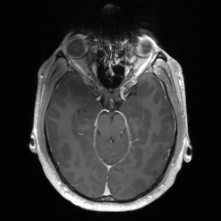
Astrocytoma_(Radiopaedia_85660-101440_I_63).jpg(247 × 247 pixels, file size: 11 KB, MIME type: image/jpeg)

Summary:

Description
  • Radiopaedia case ID: 85660
  • Image ID: 54169550
  • Image stack position: 63/180
  • Plane projection: Axial
  • Aux modality: T1 C+
  • Modality: MRI
  • System: Central Nervous System
  • Findings: Right frontal lesion has 2 components:a nodular component medially that has herniated beneath the falx to cross midline with multiple small vessels and wispy enhancement, and a crescentic lateral component that demonstrates the T2 FLAIR mismatch sign. There is additional surrounding peritumoural edema. The lesion appears intra-axial and solitary. Local mass effect has compressed the right frontal horn and created a midline shift of 1 cm and has displaced the 2 A2 branches (in close proximity to its left posterior margin)No significant diffusion restriction. No calcification on CT. No internal hemorrhage.Conclusion:Solitary right frontal intra-axial lesion is most likely to represent a glioma. The T2 FLAIR mismatch favors a non 1p19q codeleted IDH mutant diffuse astrocytoma while the more medial nodule of wispy enhancement with internal vascularity is suspicious for a high-grade/anaplastic component.
Date Published: 21st Apr 2021
Source https://radiopaedia.org/cases/astrocytoma-2
Author Frank Gaillard
Permission
(Permission-reusing-text)
http://creativecommons.org/licenses/by-nc-sa/3.0/

Licensing:

Attribution-NonCommercial-ShareAlike 3.0 Unported (CC BY-NC-SA 3.0)

File history

Click on a date/time to view the file as it appeared at that time.

Date/TimeThumbnailDimensionsUserComment
current00:18, 28 May 2021Thumbnail for version as of 00:18, 28 May 2021247 × 247 (11 KB) (talk | contribs)Radiopaedia project rID:85660 (batch #3105-868 I63)

The following page uses this file: