File:Atlanto-axial subluxation-dislocation (Radiopaedia 29659-30185 Axial bone window 20).jpg
Jump to navigation
Jump to search
Size of this preview: 636 × 600 pixels. Other resolutions: 254 × 240 pixels | 509 × 480 pixels | 740 × 698 pixels.
Original file (740 × 698 pixels, file size: 48 KB, MIME type: image/jpeg)
Summary:
| Description |
|
| Date | 12 Jun 2014 |
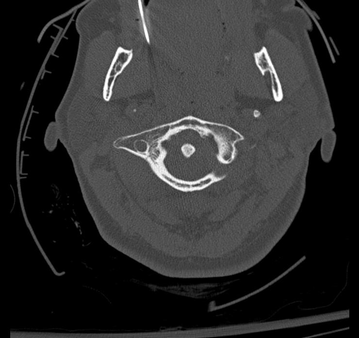
| Source | Atlanto-axial subluxation/dislocation |
| Author | Eric F Greif |
| Permission (Permission-reusing-text) |
http://creativecommons.org/licenses/by-nc-sa/3.0/ |
Licensing:
Attribution-NonCommercial-ShareAlike 3.0 Unported (CC BY-NC-SA 3.0)
| This file is ineligible for copyright and therefore in the public domain, because it is a technical image created as part of a standard medical diagnostic procedure. No creative element rising above the threshold of originality was involved in its production.
|
 |
File history
Click on a date/time to view the file as it appeared at that time.
| Date/Time | Thumbnail | Dimensions | User | Comment | |
|---|---|---|---|---|---|
| current | 21:13, 28 May 2021 | | 740 × 698 (48 KB) | Fæ (talk | contribs) | Radiopaedia project rID:29659 (batch #3138-59 C20) |
You cannot overwrite this file.
.jpg&oldid=9452624)