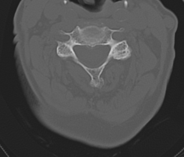
File:Atlanto-axial subluxation (Radiopaedia 36610-38176 Axial bone window 28).png
Jump to navigation
Jump to search
Atlanto-axial_subluxation_(Radiopaedia_36610-38176_Axial_bone_window_28).png (602 × 512 pixels, file size: 191 KB, MIME type: image/png)
Summary:
| Description |
|
| Date | Published: 3rd May 2015 |
| Source | https://radiopaedia.org/cases/atlanto-axial-subluxation |
| Author | Henry Knipe |
| Permission (Permission-reusing-text) |
http://creativecommons.org/licenses/by-nc-sa/3.0/ |
Licensing:
Attribution-NonCommercial-ShareAlike 3.0 Unported (CC BY-NC-SA 3.0)
File history
Click on a date/time to view the file as it appeared at that time.
| Date/Time | Thumbnail | Dimensions | User | Comment | |
|---|---|---|---|---|---|
| current | 20:03, 28 May 2021 |  | 602 × 512 (191 KB) | Fæ (talk | contribs) | Radiopaedia project rID:36610 (batch #3135-110 B28) |
You cannot overwrite this file.
File usage
The following page uses this file:
