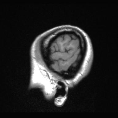
File:Atretic encephalocoele with inferior vermis hypoplasia (Radiopaedia 30443-31108 Sagittal T1 5).jpg

From NC Commons
Jump to navigation Jump to search

Summary:

Description
  • Radiopaedia case ID: 30443
  • Image ID: 7980732
  • Image stack position: 5/126
  • Plane projection: Sagittal
  • Aux modality: T1
  • Study findings: Hypoplasia of the inferior vermis, with enlargement of the fourth ventricle. The cerebellar hemispheres appear normal volume, but somewhat distorted. There is enlargement of the posterior fossa, with elevation of the torcula Herophili, which appears to be due to distortion of both the tentorium and adjacent parenchyma. A midline calvarial defect posterior to this shows extension of meninges into the subcutaneous tissue, with an associated 7 mm fat signal (high T1 and T2) lesion. No extracranial CSF communication. Despite the proximity to the torcula Herophili (immediately above the calvarial defect), there is no convincing evidence of vascular communication. Below this is a 2.5 x 2.3 x 2.3 fluid intensity cyst in the posterior fossa is relatively thick-walled, and there is no visible communication with the subarachnoid cisterns. No callosal dysgenesis. The grey-white matter junction and appearance of the cortex is unremarkable, with no evidence of cortical dysplasia or grey matter heterotopia. The anterior part of the left hippocampus is abnormal, slightly of higher T2 signal, with distorted morphology and loss of internal architecture. Otherwise the mesial temporal lobes, including the hippocampi, are unremarkable in size, signal and morphology. Conclusion: Enlargement of posterior fossa, elevation of the torcula Herophili and inferior vermian hypoplasia (consistent with a mild form of Dandy-Walker spectrum abnormality). Posterior midline calvarial defect, with extrusion of meninges, has the appearance of an atretic encephalocoele. The presence of fat signal within the extracranial portion suggests a lipomatous component. The meningeal component appears to communicate with the covering of the posterior fossa cyst, which may represent the intracranial portion of the same defect. Should intervention be considered upon these defects are then vascular imaging is recommended. Abnormal left anterior hippocampus likely dysplastic.
  • Modality: MRI
  • System: Central Nervous System
  • Findings: The posterior cranial fossa is abnormal. The inferior cerebellar vermis is  hypoplastic and there is continuity between the cisterna magna and dilated fourth ventricle. The tentorium cerebelli is high. The vermis is present, but small. Scalloping of the posterior cranial fossa inner table (remodeling). Conclusions: Findings are in keeping with a Dandy-Walker spectrum abnormality with cerebellar hemispheric and vermian hypoplasia.
Date Published: 11th Aug 2014
Source https://radiopaedia.org/cases/atretic-encephalocoele-with-inferior-vermis-hypoplasia
Author Paul Simkin
Permission
(Permission-reusing-text)
http://creativecommons.org/licenses/by-nc-sa/3.0/

Licensing:

Attribution-NonCommercial-ShareAlike 3.0 Unported (CC BY-NC-SA 3.0)

File history

Click on a date/time to view the file as it appeared at that time.

Date/TimeThumbnailDimensionsUserComment
current09:06, 29 May 2021Thumbnail for version as of 09:06, 29 May 2021384 × 384 (10 KB) (talk | contribs)Radiopaedia project rID:30443 (batch #3167-534 F5)