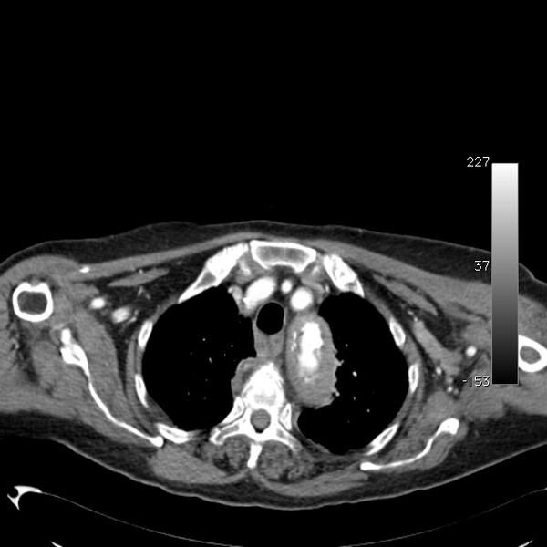
File:Atypical dissection of the thoracic aorta (Radiopaedia 10975-11393 A 9).jpg
Jump to navigation
Jump to search

Size of this preview: 600 × 600 pixels. Other resolutions: 240 × 240 pixels | 631 × 631 pixels.
Original file (631 × 631 pixels, file size: 74 KB, MIME type: image/jpeg)
Summary:
| Description |
|
| Date | Published: 5th Oct 2010 |
| Source | https://radiopaedia.org/cases/atypical-dissection-of-the-thoracic-aorta |
| Author | Erik Ranschaert |
| Permission (Permission-reusing-text) |
http://creativecommons.org/licenses/by-nc-sa/3.0/ |
Licensing:
Attribution-NonCommercial-ShareAlike 3.0 Unported (CC BY-NC-SA 3.0)
File history
Click on a date/time to view the file as it appeared at that time.
| Date/Time | Thumbnail | Dimensions | User | Comment | |
|---|---|---|---|---|---|
| current | 15:07, 29 May 2021 |  | 631 × 631 (74 KB) | Fæ (talk | contribs) | Radiopaedia project rID:10975 (batch #3211-9 A9) |
You cannot overwrite this file.
File usage
The following page uses this file: