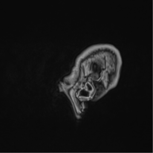
File:Atypical meningioma (WHO Grade II) (Radiopaedia 54742-60979 Sagittal T1 C+ 4).png
Jump to navigation
Jump to search
No higher resolution available.
Atypical_meningioma_(WHO_Grade_II)_(Radiopaedia_54742-60979_Sagittal_T1_C+_4).png (512 × 512 pixels, file size: 39 KB, MIME type: image/png)
Summary:
| Description |
|
| Date | Published: 30th Jul 2017 |
| Source | https://radiopaedia.org/cases/atypical-meningioma-who-grade-ii-1 |
| Author | Ernest Lekgabe |
| Permission (Permission-reusing-text) |
http://creativecommons.org/licenses/by-nc-sa/3.0/ |
Licensing:
Attribution-NonCommercial-ShareAlike 3.0 Unported (CC BY-NC-SA 3.0)
File history
Click on a date/time to view the file as it appeared at that time.
| Date/Time | Thumbnail | Dimensions | User | Comment | |
|---|---|---|---|---|---|
| current | 04:05, 3 June 2021 | | 512 × 512 (39 KB) | Fæ (talk | contribs) | Radiopaedia project rID:54742 (batch #3240-246 G4) |
You cannot overwrite this file.
File usage
The following page uses this file:
_(Radiopaedia_54742-60979_Sagittal_T1_C%2B_4).png&oldid=443274)