File:Atypical meningioma (WHO grade II) (Radiopaedia 5273-7016 Axial T2 9).jpg
Jump to navigation
Jump to search

Size of this preview: 637 × 600 pixels. Other resolutions: 255 × 240 pixels | 510 × 480 pixels | 816 × 768 pixels | 1,088 × 1,024 pixels | 1,534 × 1,444 pixels.
Original file (1,534 × 1,444 pixels, file size: 143 KB, MIME type: image/jpeg)
Summary:
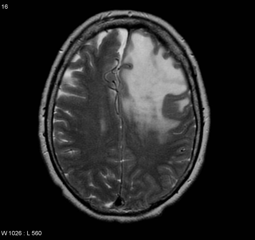
| Description |
|
| Date | Published: 29th Dec 2008 |
| Source | https://radiopaedia.org/cases/atypical-meningioma-who-grade-ii-3 |
| Author | Frank Gaillard |
| Permission (Permission-reusing-text) |
http://creativecommons.org/licenses/by-nc-sa/3.0/ |
Licensing:
Attribution-NonCommercial-ShareAlike 3.0 Unported (CC BY-NC-SA 3.0)
File history
Click on a date/time to view the file as it appeared at that time.
| Date/Time | Thumbnail | Dimensions | User | Comment | |
|---|---|---|---|---|---|
| current | 02:53, 3 June 2021 | | 1,534 × 1,444 (143 KB) | Fæ (talk | contribs) | Radiopaedia project rID:5273 (batch #3237-11 C9) |
You cannot overwrite this file.
File usage
There are no pages that use this file.
_(Radiopaedia_5273-7016_Axial_T2_9).jpg&oldid=442795)