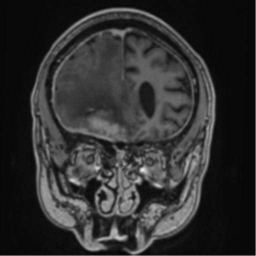
File:Atypical meningioma (WHO grade II) with brain invasion (Radiopaedia 57767-64729 Coronal T1 C+ 20).png

From NC Commons
Jump to navigation Jump to search

Summary:

Description
  • Radiopaedia case ID: 57767
  • Image ID: 35148085
  • Image stack position: 20/90
  • Plane projection: Coronal
  • Aux modality: T1 C+
  • Study description: MRI Brain
  • Study findings: Corresponding to the mass on CT, is a lobulated mass in the right frontal region, abutting inner table of the calvarium, with dural tails and moderately extensive adjacent dural thickening. Foci of internal susceptibility correspond to the calcification on CT. This lesion crosses the midline to the left, moderately indenting the left gyrus rectus. It also extends anteriorly through the inner table of the right frontal sinus with the right frontal sinus opacified by a combination of enhancing tumor and mucosa. It is uncertain whether the adjacent anterior-most ethmoidal air cell is also involved. Moderate surrounding FLAIR hyperintensity with local mass effect, including distortion of the right frontal horn and midline shift of 9 mm to the left. Interposing FLAIR hypointensity in keeping with peritumoral cysts. The mass shows high CBV and serpentine enhancement likely vessels in the adjacent brains in keeping with high vascularity. Some borders between the tumor and parenchyma are ill-defined, and the possibility of invasion needs to be considered. MRS shows elevated choline, probable glx and alanine peaks on a broadened NAA peak that merges with a broadened lactate peak. Similarly vividly enhancing left paraclinoid mass lesion is also noted, appearing to abut the left lateral optic nerves, medially displacing it, and abutting the undersurface of the left terminal carotid.
  • Modality: MRI
  • System: Central Nervous System
  • Findings: There is a large right frontal region extra-axial tumor that is solid and hyperdense, with some flecks of peripheral calcification, cause significant mass effect pushing the adjacent brain parenchyma, with subfalcine herniation. The lesion causes a permeative frontal bone erosion/hyperostosis with an extra-cranial extension into the right paranasal sinus. A small lesion with similar characteristics, but smaller, is also present adjacent to the left anterior clinoid process.
Date Published: 13th Jan 2018
Source https://radiopaedia.org/cases/atypical-meningioma-who-grade-ii-with-brain-invasion
Author Bruno Di Muzio
Permission
(Permission-reusing-text)
http://creativecommons.org/licenses/by-nc-sa/3.0/

Licensing:

Attribution-NonCommercial-ShareAlike 3.0 Unported (CC BY-NC-SA 3.0)

File history

Click on a date/time to view the file as it appeared at that time.

Date/TimeThumbnailDimensionsUserComment
current06:48, 3 June 2021Thumbnail for version as of 06:48, 3 June 2021512 × 512 (85 KB) (talk | contribs)Radiopaedia project rID:57767 (batch #3241-321 F20)

Metadata