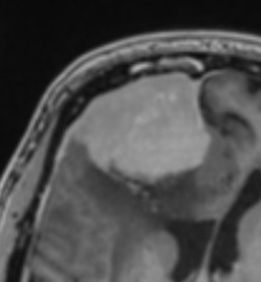
File:Atypical meningioma (WHO grade II) with brain invasion (Radiopaedia 57767-64766 T1 C+ 1).png
Jump to navigation
Jump to search
Atypical_meningioma_(WHO_grade_II)_with_brain_invasion_(Radiopaedia_57767-64766_T1_C+_1).png (513 × 554 pixels, file size: 142 KB, MIME type: image/png)
Summary:
| Description |
|
| Date | Published: 13th Jan 2018 |
| Source | https://radiopaedia.org/cases/atypical-meningioma-who-grade-ii-with-brain-invasion |
| Author | Bruno Di Muzio |
| Permission (Permission-reusing-text) |
http://creativecommons.org/licenses/by-nc-sa/3.0/ |
Licensing:
Attribution-NonCommercial-ShareAlike 3.0 Unported (CC BY-NC-SA 3.0)
File history
Click on a date/time to view the file as it appeared at that time.
| Date/Time | Thumbnail | Dimensions | User | Comment | |
|---|---|---|---|---|---|
| current | 07:30, 3 June 2021 |  | 513 × 554 (142 KB) | Fæ (talk | contribs) | Radiopaedia project rID:57767 (batch #3241-2 B1) |
You cannot overwrite this file.
File usage
There are no pages that use this file.
