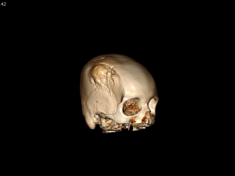
File:Atypical meningioma - intraosseous (Radiopaedia 64915-73867 3D volume render 42).jpg

From NC Commons
Jump to navigation Jump to search

Original file(1,780 × 1,334 pixels, file size: 199 KB, MIME type: image/jpeg)

Summary:

Description
  • Radiopaedia case ID: 64915
  • Study ID: 73867
  • Image ID: 45421224
  • Plane projection: 3D volume render
  • Modality: CT
  • System: Central Nervous System
  • Findings: There is an expansile lesion centered in the right parietal bone, infiltrating and expanding the diploic space. The lesion is heterogeneously T1 and T2 hypointense and has areas of abnormally restricted diffusion. The lesion measures 8 cm in maximal diameter and extends laterally into the adjacent soft tissues and medially into the epidural space. There is mild local mass effect with mild effacement of adjacent sulci, without associated increased T2/FLAIR hyperintensity in the brain parenchyma. This may represent an intraosseous meningioma or fibrous dysplasia with other etiologies also possible. Head CT is recommended to further characterize the lesion. Other findings:
  • Study findings: An expansile lytic and sclerotic bone lesion is centered in the right parietal bone. The lesion measures 8 cm in maximal diameter. The lesion extends anteriorly just beyond the coronal suture into the right frontal bone and inferiorly to the squamosal suture. The involved outer table is irregular and the inner table is spiculated. The lesion is associated with enhancing soft tissue that extends laterally about 3 mm into the adjacent scalp under the temporalis muscle and medially about 1 cm into the epidural space. There is evidence of mild mass effect with mild effacement of the sulci in the adjacent right posterior frontal lobe. There is no underlying edema pattern or midline shift. Cortical veins overlie this region and may be partially encased by the soft tissue component. Together with the long duration of symptoms, the appearance favors intraosseous meningioma, perhaps of a higher grade given the degree of bony erosion. Cavernous hemangioma is an alternative consideration but this lesion typically spares the inner table, which is involved on the current study. Fibrous dysplasia, which relatively rarely involves the calvaria, is unlikely because it typically preserves the inner and outer tables even when it causes osseous expansion. More malignant etiologies such as lymphoma and metastases, despite the restricted diffusion seen on MRI, are felt to be unlikely given the hyperostotic component and long duration of symptoms. Osteosarcoma is also unlikely given the long course. Other findings:
  • Author location: Baltimore, United States
Date 29 Jun 2019
Source Atypical meningioma - intraosseous
Author Francis Deng
Permission
(Permission-reusing-text)
http://creativecommons.org/licenses/by-nc-sa/3.0/

Licensing:

Attribution-NonCommercial-ShareAlike 3.0 Unported (CC BY-NC-SA 3.0)

This file is ineligible for copyright and therefore in the public domain, because it is a technical image created as part of a standard medical diagnostic procedure. No creative element rising above the threshold of originality was involved in its production.

File history

Click on a date/time to view the file as it appeared at that time.

Date/TimeThumbnailDimensionsUserComment
current02:31, 3 June 2021Thumbnail for version as of 02:31, 3 June 20211,780 × 1,334 (199 KB) (talk | contribs)Radiopaedia project rID:64915 (batch #3236-515 H42)