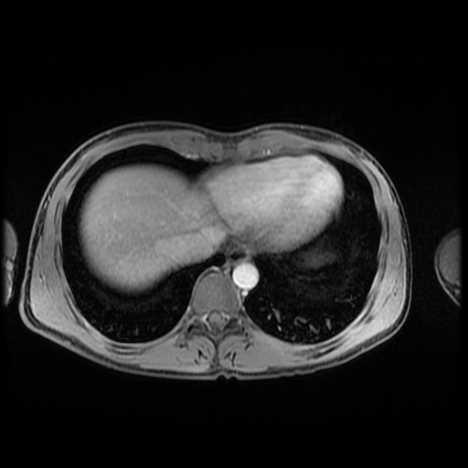
File:Autoimmune pancreatitis (Radiopaedia 69751-79729 N 10).jpg
Jump to navigation
Jump to search
Autoimmune_pancreatitis_(Radiopaedia_69751-79729_N_10).jpg (512 × 512 pixels, file size: 46 KB, MIME type: image/jpeg)
Summary:
| Description |
|
| Date | Published: 2nd Nov 2019 |
| Source | https://radiopaedia.org/cases/autoimmune-pancreatitis-9 |
| Author | Bruno Di Muzio |
| Permission (Permission-reusing-text) |
http://creativecommons.org/licenses/by-nc-sa/3.0/ |
Licensing:
Attribution-NonCommercial-ShareAlike 3.0 Unported (CC BY-NC-SA 3.0)
File history
Click on a date/time to view the file as it appeared at that time.
| Date/Time | Thumbnail | Dimensions | User | Comment | |
|---|---|---|---|---|---|
| current | 22:26, 3 June 2021 |  | 512 × 512 (46 KB) | Fæ (talk | contribs) | Radiopaedia project rID:69751 (batch #3274-728 N10) |
You cannot overwrite this file.
File usage
The following page uses this file:
