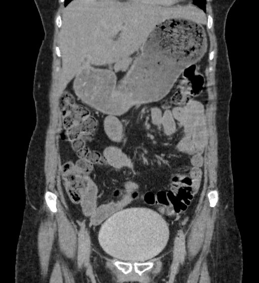
File:Autosomal dominant polycystic kidney disease (Radiopaedia 38189-40194 Coronal C+ delayed 19).jpg
Jump to navigation
Jump to search
No higher resolution available.
Autosomal_dominant_polycystic_kidney_disease_(Radiopaedia_38189-40194_Coronal_C+_delayed_19).jpg (512 × 559 pixels, file size: 150 KB, MIME type: image/jpeg)
Summary:
| Description |
|
| Date | Published: 11th Jul 2015 |
| Source | https://radiopaedia.org/cases/autosomal-dominant-polycystic-kidney-disease-15 |
| Author | Vinay TP |
| Permission (Permission-reusing-text) |
http://creativecommons.org/licenses/by-nc-sa/3.0/ |
Licensing:
Attribution-NonCommercial-ShareAlike 3.0 Unported (CC BY-NC-SA 3.0)
File history
Click on a date/time to view the file as it appeared at that time.
| Date/Time | Thumbnail | Dimensions | User | Comment | |
|---|---|---|---|---|---|
| current | 06:43, 4 June 2021 | | 512 × 559 (150 KB) | Fæ (talk | contribs) | Radiopaedia project rID:38189 (batch #3292-83 B19) |
You cannot overwrite this file.
File usage
The following page uses this file:
.jpg&oldid=453705)