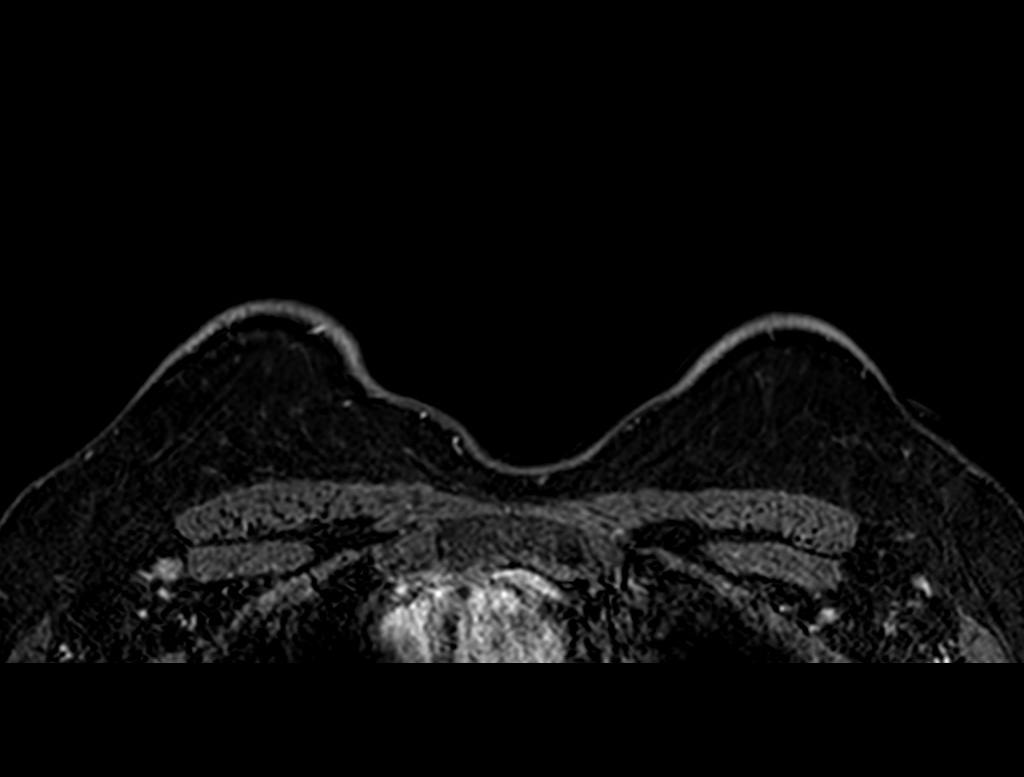
File:Axillary lymph nodes - surgical levels (Radiopaedia 84413-104719 Axial T1 C+ fat sat 76).jpg
Jump to navigation
Jump to search
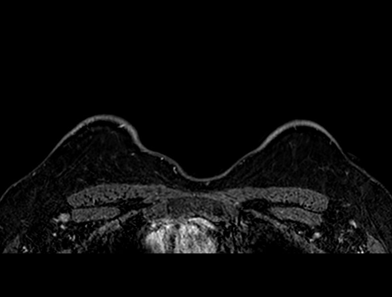
Size of this preview: 790 × 599 pixels. Other resolutions: 316 × 240 pixels | 633 × 480 pixels | 1,012 × 768 pixels | 1,024 × 777 pixels.
Original file (1,024 × 777 pixels, file size: 79 KB, MIME type: image/jpeg)
Summary:
| Description |
|
| Date | Published: 31st Mar 2021 |
| Source | https://radiopaedia.org/cases/axillary-lymph-nodes-surgical-levels |
| Author | Israel Rodriguez-Suarez |
| Permission (Permission-reusing-text) |
http://creativecommons.org/licenses/by-nc-sa/3.0/ |
Licensing:
Attribution-NonCommercial-ShareAlike 3.0 Unported (CC BY-NC-SA 3.0)
File history
Click on a date/time to view the file as it appeared at that time.
| Date/Time | Thumbnail | Dimensions | User | Comment | |
|---|---|---|---|---|---|
| current | 10:08, 5 June 2021 | | 1,024 × 777 (79 KB) | Fæ (talk | contribs) | Radiopaedia project rID:84413 (batch #3478-112 B76) |
You cannot overwrite this file.
File usage
The following page uses this file:
.jpg&oldid=464412)