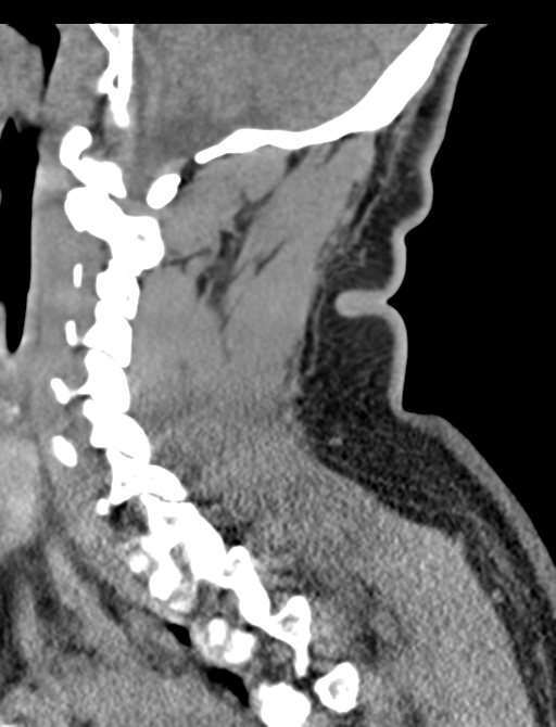
File:Axis peg fracture (type 3) and atlas lateral mass (type 4) fracture (Radiopaedia 37474-39324 D 29).png
Jump to navigation
Jump to search
Size of this preview: 458 × 599 pixels. Other resolutions: 183 × 240 pixels | 512 × 670 pixels.
Original file (512 × 670 pixels, file size: 293 KB, MIME type: image/png)
Summary:
| Description |
|
| Date | Published: 28th Jul 2015 |
| Source | https://radiopaedia.org/cases/axis-peg-fracture-type-3-and-atlas-lateral-mass-type-4-fracture |
| Author | Craig Hacking |
| Permission (Permission-reusing-text) |
http://creativecommons.org/licenses/by-nc-sa/3.0/ |
Licensing:
Attribution-NonCommercial-ShareAlike 3.0 Unported (CC BY-NC-SA 3.0)
File history
Click on a date/time to view the file as it appeared at that time.
| Date/Time | Thumbnail | Dimensions | User | Comment | |
|---|---|---|---|---|---|
| current | 11:52, 5 June 2021 | | 512 × 670 (293 KB) | Fæ (talk | contribs) | Radiopaedia project rID:37474 (batch #3487-266 D29) |
You cannot overwrite this file.
File usage
The following page uses this file:
_and_atlas_lateral_mass_(type_4)_fracture_(Radiopaedia_37474-39324_D_29).png&oldid=465063)