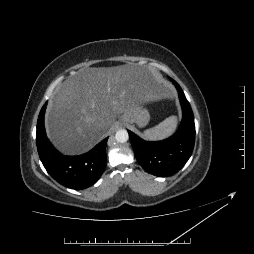
File:Azygos continuation of the IVC (Radiopaedia 40416-42965 A 11).jpg
Jump to navigation
Jump to search
Azygos_continuation_of_the_IVC_(Radiopaedia_40416-42965_A_11).jpg (512 × 512 pixels, file size: 43 KB, MIME type: image/jpeg)
Summary:
| Description |
|
| Date | Published: 22nd Apr 2016 |
| Source | https://radiopaedia.org/cases/azygos-continuation-of-the-ivc-1 |
| Author | Essam G Ghonaim |
| Permission (Permission-reusing-text) |
http://creativecommons.org/licenses/by-nc-sa/3.0/ |
Licensing:
Attribution-NonCommercial-ShareAlike 3.0 Unported (CC BY-NC-SA 3.0)
File history
Click on a date/time to view the file as it appeared at that time.
| Date/Time | Thumbnail | Dimensions | User | Comment | |
|---|---|---|---|---|---|
| current | 17:06, 5 June 2021 |  | 512 × 512 (43 KB) | Fæ (talk | contribs) | Radiopaedia project rID:40416 (batch #3510-11 A11) |
You cannot overwrite this file.
File usage
There are no pages that use this file.
