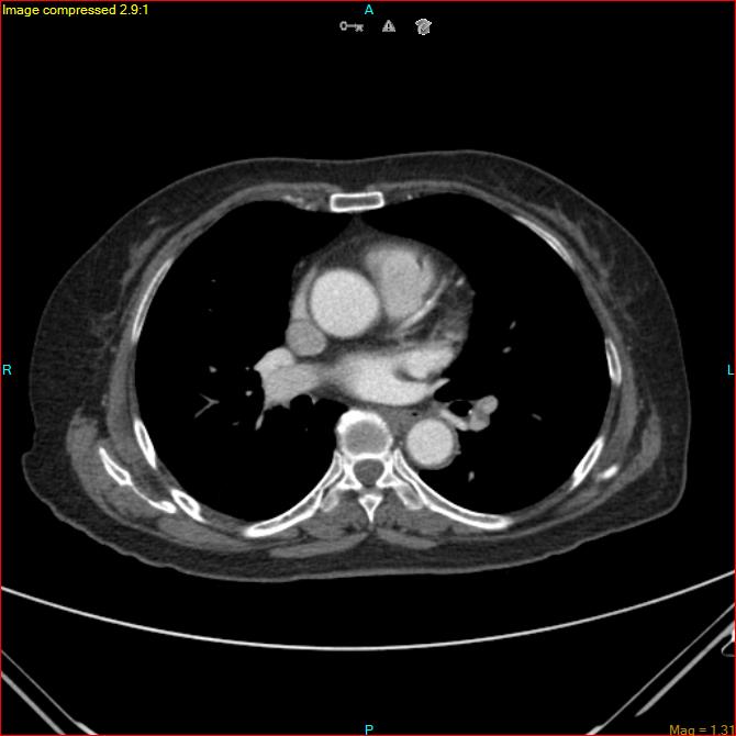
File:Azygos vein aneurysm (Radiopaedia 77824-90130 B 31).jpg
Jump to navigation
Jump to search
Size of this preview: 600 × 600 pixels. Other resolutions: 240 × 240 pixels | 480 × 480 pixels | 670 × 670 pixels.
Original file (670 × 670 pixels, file size: 41 KB, MIME type: image/jpeg)
Summary:
| Description |
|
| Date | Published: 27th May 2020 |
| Source | https://radiopaedia.org/cases/azygos-vein-aneurysm |
| Author | Anton Lorenzo Gutierrez |
| Permission (Permission-reusing-text) |
http://creativecommons.org/licenses/by-nc-sa/3.0/ |
Licensing:
Attribution-NonCommercial-ShareAlike 3.0 Unported (CC BY-NC-SA 3.0)
File history
Click on a date/time to view the file as it appeared at that time.
| Date/Time | Thumbnail | Dimensions | User | Comment | |
|---|---|---|---|---|---|
| current | 19:43, 5 June 2021 | | 670 × 670 (41 KB) | Fæ (talk | contribs) | Radiopaedia project rID:77824 (batch #3548-86 B31) |
You cannot overwrite this file.
File usage
The following page uses this file:
.jpg&oldid=468119)