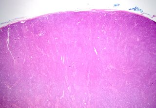
File:Basal cell adenoma - salivary gland (Radiopaedia 43989-47511 20x H&E 1).jpg
Jump to navigation
Jump to search

Size of this preview: 800 × 558 pixels. Other resolutions: 320 × 223 pixels | 640 × 446 pixels | 1,024 × 714 pixels | 1,280 × 892 pixels | 2,517 × 1,755 pixels.
Original file (2,517 × 1,755 pixels, file size: 959 KB, MIME type: image/jpeg)
Summary:
| Description |
|
| Date | Published: 1st Apr 2016 |
| Source | https://radiopaedia.org/cases/basal-cell-adenoma-salivary-gland-1 |
| Author | Mikkaela McCormack |
| Permission (Permission-reusing-text) |
http://creativecommons.org/licenses/by-nc-sa/3.0/ |
Licensing:
Attribution-NonCommercial-ShareAlike 3.0 Unported (CC BY-NC-SA 3.0)
File history
Click on a date/time to view the file as it appeared at that time.
| Date/Time | Thumbnail | Dimensions | User | Comment | |
|---|---|---|---|---|---|
| current | 23:59, 6 June 2021 | | 2,517 × 1,755 (959 KB) | Fæ (talk | contribs) | Radiopaedia project rID:43989 (batch #3660-1 A1) |
You cannot overwrite this file.
File usage
There are no pages that use this file.
.jpg&oldid=478859)