File:Basal ganglia (illustration) (Radiopaedia 36285).jpg
Jump to navigation
Jump to search
No higher resolution available.
Basal_ganglia_(illustration)_(Radiopaedia_36285).jpg (800 × 350 pixels, file size: 57 KB, MIME type: image/jpeg)
Summary:
- Radiopaedia case ID: 36285
- Image ID: 23674
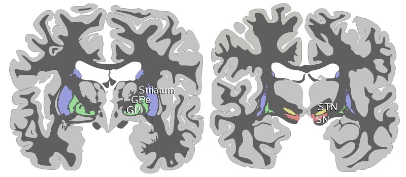
- Study findings: Two schematic drawings of coronal sections of human brain labeling the basal ganglia. Blue=striatum, green=globus pallidus (external and internal segments), yellow=subthalamic nucleus, red=substantia nigra (pars reticulata and pars compacta). The right section is the deeper one, closer to the back of the head. Images taken from wikipedia from the out of copyright Gray's anatomy. Basal ganglia locationOriginal file: http://commons.wikimedia.org/wiki/File:Telencephalon-Horiconatal.jpgBasal ganglia (coronal)Permission: GFDLOriginal file: http://commons.wikimedia.org/wiki/File:Basal-ganglia-coronal-sections-large.pngDopamine in PDThe image shows dopaminergic pathways of the human brain in normal condition (left) and Parkinsons Disease (right). Red Arrows indicate suppression of the target, blue arrows indicate stimulation of target structure.Permission: GNU freeOriginal file: http://commons.wikimedia.org/wiki/File:DA-loops_in_PD.jpg
- Modality: Diagram
- System: Central Nervous System
- Findings: Two schematic drawings of coronal sections of human brain labeling the basal ganglia. Blue=striatum, green=globus pallidus (external and internal segments), yellow=subthalamic nucleus, red=substantia nigra (pars reticulata and pars compacta). The right section is the deeper one, closer to the back of the head. Images taken from wikipedia from the out of copyright Gray's anatomy. Basal ganglia locationOriginal file: http: //commons. wikimedia. org/wiki/File: Telencephalon-Horiconatal. jpgBasal ganglia (coronal)Permission: GFDLOriginal file: http: //commons. wikimedia. org/wiki/File: Basal-ganglia-coronal-sections-large. pngDopamine in PDThe image shows dopaminergic pathways of the human brain in normal condition (left) and Parkinsons Disease (right). Red Arrows indicate suppression of the target, blue arrows indicate stimulation of target structure. Permission: GNU freeOriginal file: http: //commons. wikimedia. org/wiki/File: DA-loops_in_PD. jpg
- Published: 16th May 2015
- Source: https://radiopaedia.org/cases/basal-ganglia-illustration
- Author: Wikipedia
- Permission: http://creativecommons.org/licenses/by-nc-sa/3.0/
Licensing:
Attribution-NonCommercial-ShareAlike 3.0 Unported (CC BY-NC-SA 3.0)
File history
Click on a date/time to view the file as it appeared at that time.
| Date/Time | Thumbnail | Dimensions | User | Comment | |
|---|---|---|---|---|---|
| current | 11:42, 19 March 2021 | | 800 × 350 (57 KB) | Fæ (talk | contribs) | Radiopaedia project rID:36285 (batch #3597) |
You cannot overwrite this file.
File usage
The following page uses this file:
_(Radiopaedia_36285).jpg&oldid=8859687)