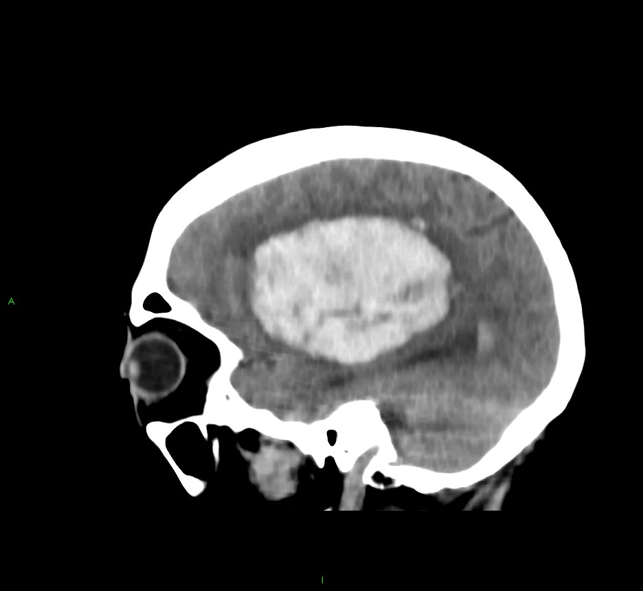
File:Basal ganglia hemorrhage (Radiopaedia 58767-65989 C 37).jpg
Jump to navigation
Jump to search
Size of this preview: 653 × 600 pixels. Other resolutions: 261 × 240 pixels | 522 × 480 pixels | 936 × 860 pixels.
Original file (936 × 860 pixels, file size: 59 KB, MIME type: image/jpeg)
Summary:
| Description |
|
| Date | Published: 6th Mar 2018 |
| Source | https://radiopaedia.org/cases/basal-ganglia-haemorrhage-9 |
| Author | Mark Rodrigues |
| Permission (Permission-reusing-text) |
http://creativecommons.org/licenses/by-nc-sa/3.0/ |
Licensing:
Attribution-NonCommercial-ShareAlike 3.0 Unported (CC BY-NC-SA 3.0)
File history
Click on a date/time to view the file as it appeared at that time.
| Date/Time | Thumbnail | Dimensions | User | Comment | |
|---|---|---|---|---|---|
| current | 03:53, 7 June 2021 | | 936 × 860 (59 KB) | Fæ (talk | contribs) | Radiopaedia project rID:58767 (batch #3677-157 C37) |
You cannot overwrite this file.
File usage
The following page uses this file:
.jpg&oldid=480404)