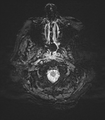
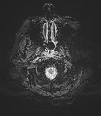
File:Basal ganglia metastasis (Radiopaedia 78928-91828 Axial SWI 2).png

From NC Commons
Jump to navigation Jump to search

Basal_ganglia_metastasis_(Radiopaedia_78928-91828_Axial_SWI_2).png(336 × 384 pixels, file size: 116 KB, MIME type: image/png)

Summary:

Description
  • Radiopaedia case ID: 78928
  • Image ID: 52926071
  • Image stack position: 2/88
  • Plane projection: Axial
  • Aux modality: SWI
  • Study findings: Within the right basal ganglia, there is a mass which demonstrates predominantly peripheral nodular contrast enhancement. This is embedded within a region of expansile FLAIR/T2 hyperintensity involving the right basal ganglia and thalamus extending into the right cerebral peduncle and right side of the midbrain. There is further extensive surrounding high T2/FLAIR signal within the white matter of the right frontoparietal region and right temporal lobe, in keeping with vasogenic edema. Right frontoparietal sulcal effacement, partial effacement of the right lateral ventricle with shift of the midline to the left. There is dilation of the left temporal horn associated with periventricular FLAIR hyperintensity suggestive of hydrocephalus with transependymal flow of CSF. The enhancing wall of the mass demonstrates low ADC and markedly elevated CBV. No central restricted diffusion. Small foci of susceptibility associated with lesion may represent blood products. MR spectroscopy demonstrates reversal of the choline/creatine ratio, depressed NAA and elevated lactate. A small region of encephalomalacia and gliosis in the left superior frontal gyrus. No other lesions demonstrated. Partial empty sella noted. Incidental developmental venous anomaly in the right hemipons. Conclusion: Right basal ganglia expansile mass with nodular contrast enhancement does not represent a pyogenic abscess. Atypical infection (especially toxoplasmosis) is a possibility. Glioblastoma may also appear similar. In the setting of the CT chest abnormalities (see later) the most likely diagnosis is, however, a metastasis.
  • Modality: MRI
  • System: Central Nervous System
  • Findings: Peripherally enhancing right basal ganglia lesion with extensive surrounding vasogenic edema.
Date Published: 18th Jun 2020
Source https://radiopaedia.org/cases/basal-ganglia-metastasis
Author Frank Gaillard
Permission
(Permission-reusing-text)
http://creativecommons.org/licenses/by-nc-sa/3.0/

Licensing:

Attribution-NonCommercial-ShareAlike 3.0 Unported (CC BY-NC-SA 3.0)

File history

Click on a date/time to view the file as it appeared at that time.

Date/TimeThumbnailDimensionsUserComment
current15:14, 7 June 2021Thumbnail for version as of 15:14, 7 June 2021336 × 384 (116 KB) (talk | contribs)Radiopaedia project rID:78928 (batch #3699-214 F2)

Metadata