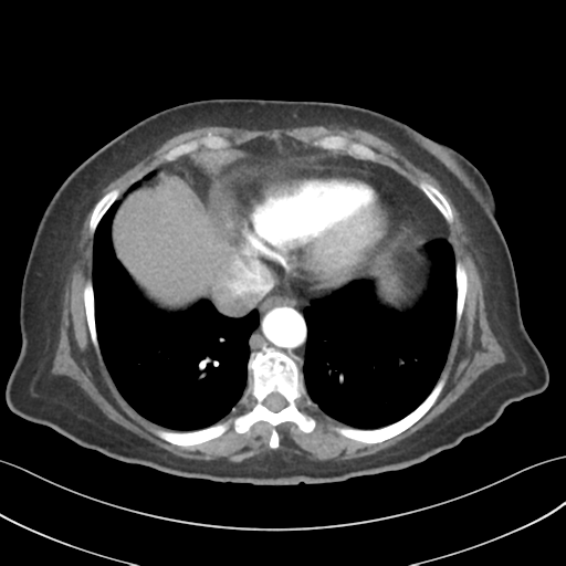
File:Basal ganglia metastasis (Radiopaedia 78928-91832 A 36).png
Jump to navigation
Jump to search
Basal_ganglia_metastasis_(Radiopaedia_78928-91832_A_36).png (512 × 512 pixels, file size: 141 KB, MIME type: image/png)
Summary:
| Description |
|
| Date | Published: 18th Jun 2020 |
| Source | https://radiopaedia.org/cases/basal-ganglia-metastasis |
| Author | Frank Gaillard |
| Permission (Permission-reusing-text) |
http://creativecommons.org/licenses/by-nc-sa/3.0/ |
Licensing:
Attribution-NonCommercial-ShareAlike 3.0 Unported (CC BY-NC-SA 3.0)
File history
Click on a date/time to view the file as it appeared at that time.
| Date/Time | Thumbnail | Dimensions | User | Comment | |
|---|---|---|---|---|---|
| current | 16:03, 7 June 2021 |  | 512 × 512 (141 KB) | Fæ (talk | contribs) | Radiopaedia project rID:78928 (batch #3699-36 A36) |
You cannot overwrite this file.
File usage
The following file is a duplicate of this file (more details):
The following page uses this file:
