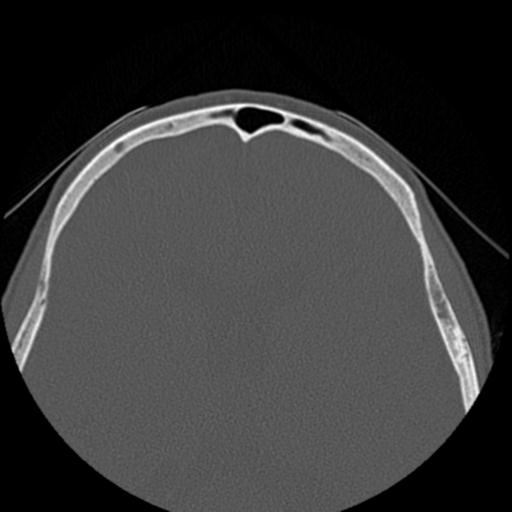
File:Base of skull chondrosarcoma (Radiopaedia 30410-31071 Axial bone window 44).jpg

From NC Commons
Jump to navigation Jump to search

Base_of_skull_chondrosarcoma_(Radiopaedia_30410-31071_Axial_bone_window_44).jpg(512 × 512 pixels, file size: 23 KB, MIME type: image/jpeg)

Summary:

Description
  • Radiopaedia case ID: 30410
  • Image ID: 7959397
  • Image stack position: 44/48
  • Plane projection: Axial
  • Aux modality: bone window
  • Study findings: In the right petrous apex a destructive lesion is present with irregular bony margins, extending to the carotid canal. It does not have a sclerotic margin nor does it have internal calcification / mineralization. Note is made of inspisated calcified right maxillary antral secretions.
  • Modality: CT
  • System: Central Nervous System
  • Findings: At the right petrous apex a soft tissue mass with intermediate T1 signal, very high T2 signal and prominent contrast enhancement, is noted. It extends to the anterior margin of the internal acoustic meatus posteriorly and to the carotid canal anteriorly. Normal flow void in the artery is preserved. No internal calcification. Conclusion: In this location, a lesion with very high T2 signal most likely represents a chondrosarcoma. Chordomas are usually more medial (midline). Other lesions would not be expected to be this enhancing and high T2 in signal (with the exception of a hemangioma, which would not be expected to have so much bony destruction).
Date Published: 11th Jul 2016
Source https://radiopaedia.org/cases/base-of-skull-chondrosarcoma-1
Author Frank Gaillard
Permission
(Permission-reusing-text)
http://creativecommons.org/licenses/by-nc-sa/3.0/

Licensing:

Attribution-NonCommercial-ShareAlike 3.0 Unported (CC BY-NC-SA 3.0)

File history

Click on a date/time to view the file as it appeared at that time.

Date/TimeThumbnailDimensionsUserComment
current21:35, 7 June 2021Thumbnail for version as of 21:35, 7 June 2021512 × 512 (23 KB) (talk | contribs)Radiopaedia project rID:30410 (batch #3730-44 A44)

There are no pages that use this file.