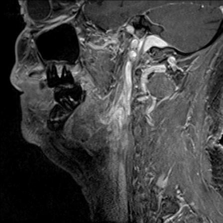
File:Base of tongue squamous cell carcinoma (Radiopaedia 31174-31884 F 123).jpg
Jump to navigation
Jump to search
Base_of_tongue_squamous_cell_carcinoma_(Radiopaedia_31174-31884_F_123).jpg (448 × 448 pixels, file size: 28 KB, MIME type: image/jpeg)
Summary:
| Description |
|
| Date | Published: 25th Feb 2015 |
| Source | https://radiopaedia.org/cases/base-of-tongue-squamous-cell-carcinoma-1 |
| Author | Smita Deb |
| Permission (Permission-reusing-text) |
http://creativecommons.org/licenses/by-nc-sa/3.0/ |
Licensing:
Attribution-NonCommercial-ShareAlike 3.0 Unported (CC BY-NC-SA 3.0)
File history
Click on a date/time to view the file as it appeared at that time.
| Date/Time | Thumbnail | Dimensions | User | Comment | |
|---|---|---|---|---|---|
| current | 00:28, 8 June 2021 |  | 448 × 448 (28 KB) | Fæ (talk | contribs) | Radiopaedia project rID:31174 (batch #3740-270 F123) |
You cannot overwrite this file.
File usage
The following page uses this file:
