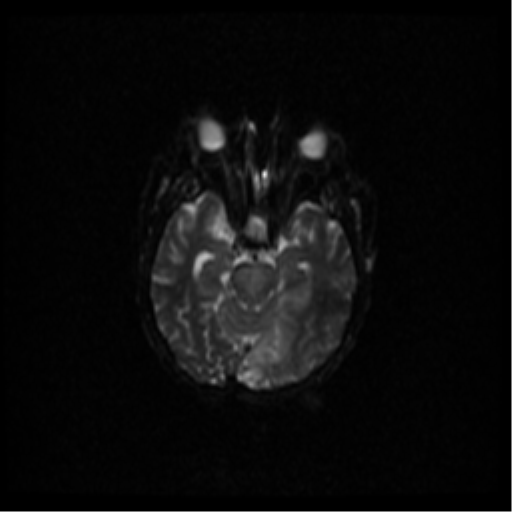
File:Basilar artery occlusion (Radiopaedia 33570-34667 Axial DWI 51).png
Jump to navigation
Jump to search
Basilar_artery_occlusion_(Radiopaedia_33570-34667_Axial_DWI_51).png (512 × 512 pixels, file size: 129 KB, MIME type: image/png)
Summary:
| Description |
|
| Date | Published: 22nd Feb 2015 |
| Source | https://radiopaedia.org/cases/basilar-artery-occlusion |
| Author | Peter Mitchell |
| Permission (Permission-reusing-text) |
http://creativecommons.org/licenses/by-nc-sa/3.0/ |
Licensing:
Attribution-NonCommercial-ShareAlike 3.0 Unported (CC BY-NC-SA 3.0)
File history
Click on a date/time to view the file as it appeared at that time.
| Date/Time | Thumbnail | Dimensions | User | Comment | |
|---|---|---|---|---|---|
| current | 06:49, 8 June 2021 |  | 512 × 512 (129 KB) | Fæ (talk | contribs) | Radiopaedia project rID:33570 (batch #3758-51 A51) |
You cannot overwrite this file.
File usage
The following page uses this file:
