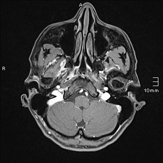
File:Basilar artery perforator aneurysm (Radiopaedia 82455-99523 G 11).jpg
Jump to navigation
Jump to search
Basilar_artery_perforator_aneurysm_(Radiopaedia_82455-99523_G_11).jpg (320 × 320 pixels, file size: 74 KB, MIME type: image/jpeg)
Summary:
| Description |
|
| Date | Published: 19th Oct 2020 |
| Source | https://radiopaedia.org/cases/basilar-artery-perforator-aneurysm |
| Author | Yves Leonard Voss |
| Permission (Permission-reusing-text) |
http://creativecommons.org/licenses/by-nc-sa/3.0/ |
Licensing:
Attribution-NonCommercial-ShareAlike 3.0 Unported (CC BY-NC-SA 3.0)
File history
Click on a date/time to view the file as it appeared at that time.
| Date/Time | Thumbnail | Dimensions | User | Comment | |
|---|---|---|---|---|---|
| current | 10:12, 8 June 2021 |  | 320 × 320 (74 KB) | Fæ (talk | contribs) | Radiopaedia project rID:82455 (batch #3760-254 G11) |
You cannot overwrite this file.
File usage
The following page uses this file:
