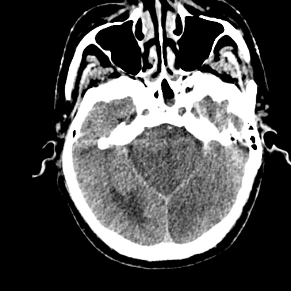
File:Basilar artery thrombosis (Radiopaedia 53351-59352 Axial non-contrast 42).jpg
Jump to navigation
Jump to search
Basilar_artery_thrombosis_(Radiopaedia_53351-59352_Axial_non-contrast_42).jpg (588 × 588 pixels, file size: 86 KB, MIME type: image/jpeg)
Summary:
| Description |
|
| Date | Published: 15th May 2017 |
| Source | https://radiopaedia.org/cases/basilar-artery-thrombosis-6 |
| Author | Ian Bickle |
| Permission (Permission-reusing-text) |
http://creativecommons.org/licenses/by-nc-sa/3.0/ |
Licensing:
Attribution-NonCommercial-ShareAlike 3.0 Unported (CC BY-NC-SA 3.0)
File history
Click on a date/time to view the file as it appeared at that time.
| Date/Time | Thumbnail | Dimensions | User | Comment | |
|---|---|---|---|---|---|
| current | 11:31, 8 June 2021 |  | 588 × 588 (86 KB) | Fæ (talk | contribs) | Radiopaedia project rID:53351 (batch #3764-42 A42) |
You cannot overwrite this file.
File usage
The following page uses this file:
