File:Benign breast clustered microcalcifications (Radiopaedia 84548-99948 B 1).png
Jump to navigation
Jump to search
Size of this preview: 731 × 600 pixels. Other resolutions: 293 × 240 pixels | 585 × 480 pixels | 936 × 768 pixels | 1,248 × 1,024 pixels | 1,820 × 1,493 pixels.
Original file (1,820 × 1,493 pixels, file size: 2.2 MB, MIME type: image/png)
Summary:
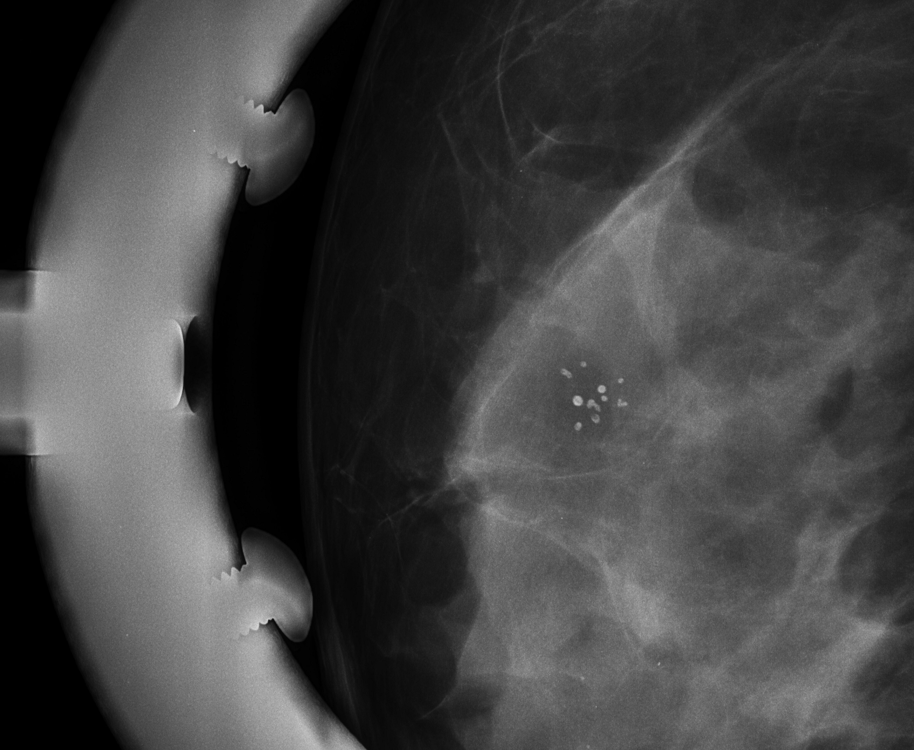
| Description |
|
| Date | Published: 4th Dec 2020 |
| Source | https://radiopaedia.org/cases/benign-breast-clustered-microcalcifications |
| Author | Mellanie Deborah |
| Permission (Permission-reusing-text) |
http://creativecommons.org/licenses/by-nc-sa/3.0/ |
Licensing:
Attribution-NonCommercial-ShareAlike 3.0 Unported (CC BY-NC-SA 3.0)
File history
Click on a date/time to view the file as it appeared at that time.
| Date/Time | Thumbnail | Dimensions | User | Comment | |
|---|---|---|---|---|---|
| current | 13:53, 9 June 2021 | | 1,820 × 1,493 (2.2 MB) | Fæ (talk | contribs) | Radiopaedia project rID:84548 (batch #3850-2 B1) |
You cannot overwrite this file.
File usage
There are no pages that use this file.
.png&oldid=502612)