File:Benign leiomyoma with hydropic features (Radiopaedia 89250-106130 A 58).jpg

From NC Commons
Jump to navigation Jump to search

Benign_leiomyoma_with_hydropic_features_(Radiopaedia_89250-106130_A_58).jpg(512 × 512 pixels, file size: 69 KB, MIME type: image/jpeg)

Summary:

Description
  • Radiopaedia case ID: 89250
  • Image ID: 54964019
  • Image stack position: 58/164
  • Plane projection: Axial
  • Aux modality: C+ portal venous phase
  • Modality: CT
  • System: Gynaecology
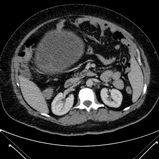
  • Findings: A large complex solid cystic mass was seen arising from the pelvis and extending into the abdomen.  2 differentials were considered most likely: 1. primary ovarian neoplastic lesion (because it appeared to arise from the right adnexa and the right ovary is not seen separately. 2. lesion arising from degeneration or neoplasia of the broad ligament fibroid (because the ovarian vein was not seen to be enlarged, the uterus appeared to possibly infiltrate the right lateral uterine wall, and prior ultrasound performed 4 years prior (see below) showed a pedunculated /broad ligament fibroid that is not seen separately on the current scan). A discrete smaller cystic lesion was seen along the left posterolateral surface of the lesion. There were a few prominent para-aortic lymph nodes of uncertain clinical significance but no definite metastatic disease in the abdominal cavity.
Date Published: 15th May 2021
Source https://radiopaedia.org/cases/benign-leiomyoma-with-hydropic-features
Author Giles Kisby
Permission
(Permission-reusing-text)
http://creativecommons.org/licenses/by-nc-sa/3.0/

Licensing:

Attribution-NonCommercial-ShareAlike 3.0 Unported (CC BY-NC-SA 3.0)

File history

Click on a date/time to view the file as it appeared at that time.

Date/TimeThumbnailDimensionsUserComment
current23:18, 9 June 2021Thumbnail for version as of 23:18, 9 June 2021512 × 512 (69 KB) (talk | contribs)Radiopaedia project rID:89250 (batch #3871-58 A58)