File:Benign mixed salivary tumor of the parotid gland (Radiopaedia 35261-36792 Axial bone window 5).jpg
Jump to navigation
Jump to search

Size of this preview: 800 × 426 pixels. Other resolutions: 320 × 170 pixels | 640 × 341 pixels | 1,184 × 630 pixels.
Original file (1,184 × 630 pixels, file size: 118 KB, MIME type: image/jpeg)
Summary:
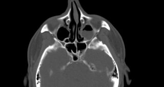
| Description |
|
| Date | Published: 29th Mar 2015 |
| Source | https://radiopaedia.org/cases/benign-mixed-salivary-tumour-of-the-parotid-gland |
| Author | Ahmed Abdrabou |
| Permission (Permission-reusing-text) |
http://creativecommons.org/licenses/by-nc-sa/3.0/ |
Licensing:
Attribution-NonCommercial-ShareAlike 3.0 Unported (CC BY-NC-SA 3.0)
File history
Click on a date/time to view the file as it appeared at that time.
| Date/Time | Thumbnail | Dimensions | User | Comment | |
|---|---|---|---|---|---|
| current | 01:03, 10 June 2021 | | 1,184 × 630 (118 KB) | Fæ (talk | contribs) | Radiopaedia project rID:35261 (batch #3875-89 D5) |
You cannot overwrite this file.
File usage
There are no pages that use this file.
.jpg&oldid=504293)