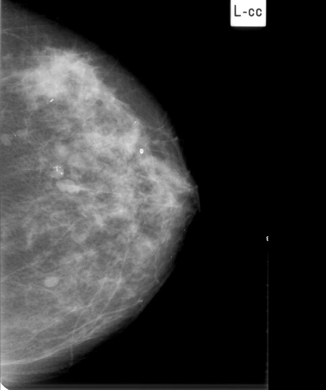
File:Benign phylloides tumor (Radiopaedia 33698-34840 CC 1).jpg
Jump to navigation
Jump to search

Size of this preview: 500 × 599 pixels. Other resolutions: 200 × 240 pixels | 401 × 480 pixels | 641 × 768 pixels | 855 × 1,024 pixels | 2,046 × 2,451 pixels.
Original file (2,046 × 2,451 pixels, file size: 818 KB, MIME type: image/jpeg)
Summary:
| Description |
|
| Date | Published: 11th Aug 2018 |
| Source | https://radiopaedia.org/cases/benign-phylloides-tumour |
| Author | Alexandra Stanislavsky |
| Permission (Permission-reusing-text) |
http://creativecommons.org/licenses/by-nc-sa/3.0/ |
Licensing:
Attribution-NonCommercial-ShareAlike 3.0 Unported (CC BY-NC-SA 3.0)
File history
Click on a date/time to view the file as it appeared at that time.
| Date/Time | Thumbnail | Dimensions | User | Comment | |
|---|---|---|---|---|---|
| current | 02:44, 10 June 2021 | | 2,046 × 2,451 (818 KB) | Fæ (talk | contribs) | Radiopaedia project rID:33698 (batch #3883-2 B1) |
You cannot overwrite this file.
File usage
There are no pages that use this file.
.jpg&oldid=504954)