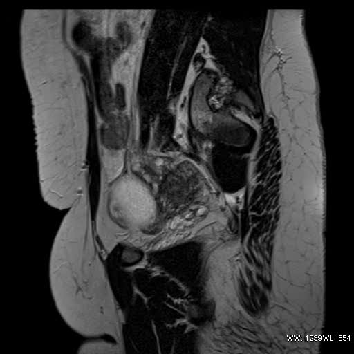
File:Bicornuate uterus- on MRI (Radiopaedia 49206-54297 Sagittal T2 22).jpg
Jump to navigation
Jump to search
Bicornuate_uterus-_on_MRI_(Radiopaedia_49206-54297_Sagittal_T2_22).jpg (512 × 512 pixels, file size: 23 KB, MIME type: image/jpeg)
Summary:
| Description |
|
| Date | Published: 15th Nov 2016 |
| Source | https://radiopaedia.org/cases/bicornuate-uterus-on-mri |
| Author | Shailaja Muniraj |
| Permission (Permission-reusing-text) |
http://creativecommons.org/licenses/by-nc-sa/3.0/ |
Licensing:
Attribution-NonCommercial-ShareAlike 3.0 Unported (CC BY-NC-SA 3.0)
File history
Click on a date/time to view the file as it appeared at that time.
| Date/Time | Thumbnail | Dimensions | User | Comment | |
|---|---|---|---|---|---|
| current | 18:27, 10 June 2021 |  | 512 × 512 (23 KB) | Fæ (talk | contribs) | Radiopaedia project rID:49206 (batch #3982-107 E22) |
You cannot overwrite this file.
File usage
There are no pages that use this file.
