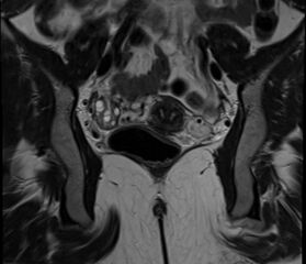
File:Bicornuate uterus (Radiopaedia 71214-81511 Coronal T2 14).jpg
Jump to navigation
Jump to search

Size of this preview: 696 × 600 pixels. Other resolutions: 279 × 240 pixels | 557 × 480 pixels | 717 × 618 pixels.
Original file (717 × 618 pixels, file size: 159 KB, MIME type: image/jpeg)
Summary:
| Description |
|
| Date | Published: 18th Oct 2019 |
| Source | https://radiopaedia.org/cases/bicornuate-uterus-22 |
| Author | Mostafa El-Feky |
| Permission (Permission-reusing-text) |
http://creativecommons.org/licenses/by-nc-sa/3.0/ |
Licensing:
Attribution-NonCommercial-ShareAlike 3.0 Unported (CC BY-NC-SA 3.0)
File history
Click on a date/time to view the file as it appeared at that time.
| Date/Time | Thumbnail | Dimensions | User | Comment | |
|---|---|---|---|---|---|
| current | 16:32, 10 June 2021 | | 717 × 618 (159 KB) | Fæ (talk | contribs) | Radiopaedia project rID:71214 (batch #3972-72 C14) |
You cannot overwrite this file.
File usage
There are no pages that use this file.
.jpg&oldid=510192)