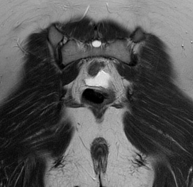
File:Bicornuate uterus (Radiopaedia 76407-88114 Coronal T2 20).jpg
Jump to navigation
Jump to search

Size of this preview: 621 × 600 pixels. Other resolutions: 249 × 240 pixels | 497 × 480 pixels | 789 × 762 pixels.
Original file (789 × 762 pixels, file size: 211 KB, MIME type: image/jpeg)
Summary:
| Description |
|
| Date | Published: 22nd Apr 2020 |
| Source | https://radiopaedia.org/cases/bicornuate-uterus-24 |
| Author | Dr Ammar Haouimi |
| Permission (Permission-reusing-text) |
http://creativecommons.org/licenses/by-nc-sa/3.0/ |
Licensing:
Attribution-NonCommercial-ShareAlike 3.0 Unported (CC BY-NC-SA 3.0)
File history
Click on a date/time to view the file as it appeared at that time.
| Date/Time | Thumbnail | Dimensions | User | Comment | |
|---|---|---|---|---|---|
| current | 15:54, 10 June 2021 |  | 789 × 762 (211 KB) | Fæ (talk | contribs) | Radiopaedia project rID:76407 (batch #3967-46 B20) |
You cannot overwrite this file.
File usage
There are no pages that use this file.