File:Bicornuate uterus (gross pathology) (Radiopaedia 9718).jpg
Jump to navigation
Jump to search

Size of this preview: 600 × 600 pixels. Other resolutions: 240 × 240 pixels | 480 × 480 pixels.
Original file (800 × 800 pixels, file size: 82 KB, MIME type: image/jpeg)
Summary:
- Radiopaedia case ID: 9718
- Image ID: 10108
- Study description: Surgical specimen
- Modality: Pathology
- System: Gynaecology
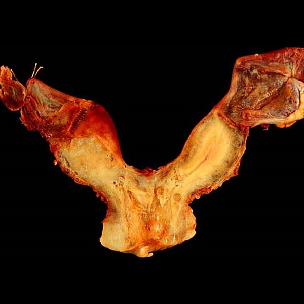
- Findings: The specimen has been cut in a coronal plane and shows features of bicornuate uterus; the proximal (lower) segment of the uterus is relatively normally formed while the distal (upper) aspect is split into two 'horns', each with it's own endometrial cavity (best seen on the right side of the photograph). Adnexal structures are attached . Image courtesy of Ed Uthman. Please see case description page for licence and original file information.
- Published: 15th May 2010
- Source: https://radiopaedia.org/cases/bicornuate-uterus-gross-pathology-3
- Author: Ed Uthman
- Permission: http://creativecommons.org/licenses/by-nc-sa/3.0/
Licensing:
Attribution-NonCommercial-ShareAlike 3.0 Unported (CC BY-NC-SA 3.0)
| This file is ineligible for copyright and therefore in the public domain, because it is a technical image created as part of a standard medical diagnostic procedure. No creative element rising above the threshold of originality was involved in its production.
|
 |
File history
Click on a date/time to view the file as it appeared at that time.
| Date/Time | Thumbnail | Dimensions | User | Comment | |
|---|---|---|---|---|---|
| current | 17:38, 19 March 2021 | | 800 × 800 (82 KB) | Fæ (talk | contribs) | Radiopaedia project rID:9718 (batch #3871) |
You cannot overwrite this file.
File usage
There are no pages that use this file.
_(Radiopaedia_9718).jpg&oldid=9755279)