File:Bilateral ICA occlusion (Radiopaedia 26044-26197 A 77).jpg

Original file (2,048 × 2,048 pixels, file size: 187 KB, MIME type: image/jpeg)
Summary:
| Description |
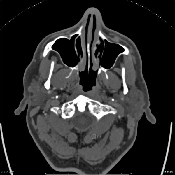
defect at the CCA bifurcation with no contrast opacification demonstrated within the cervical and virtually entire intracranial right ICA, (with only a very small amount of contrast in the supraclinoid ICA via reflux from the right posterior communicating artery). The right ECA is normal in appearance. There is contrast opacification of the proximal left CCA but no opacification of the mid and distal CCA, cervical ICA and virtually entire intracranial ICA (with only a very small amount of contrast in the distal cavernous ICA and beyond via reflux from the left posterior communicating artery) . The left ECA demonstrates contrast enhancement, (via the thyrocervical trunk). There is marked calcification of the CCA and proximal ICA bilaterally with severe luminal narrowing (L>R). Heavily calcified cavernous ICAs also noted. The right vertebral artery is dominant and demonstrates a small amount of calcification without significant luminal narrowing. Normal contrast opacification of the posterior circulation is demonstrated. There is normal contrast opacification of the remainder of the circle of willis and intracranial vasculature via the prominent posterior communicating arteries. The brachiocephalic trunk and left common carotid artery share a common origin. Conclusion no CTA evidence of flow within the cervical portions of the internal carotid arteries bilaterally, the occlusion on the left occurring at the level of the common carotid artery. Catheter angiography would be necessary to diagnose trickle flow. Normal blood flow within the circle of willis and remaining intracranial vasculature via the posterior circulation.
|
| Date | Published: 25th Nov 2013 |
| Source | https://radiopaedia.org/cases/bilateral-ica-occlusion-1 |
| Author | James Sheldon |
| Permission (Permission-reusing-text) |
http://creativecommons.org/licenses/by-nc-sa/3.0/ |
Licensing:
Attribution-NonCommercial-ShareAlike 3.0 Unported (CC BY-NC-SA 3.0)
File history
Click on a date/time to view the file as it appeared at that time.
| Date/Time | Thumbnail | Dimensions | User | Comment | |
|---|---|---|---|---|---|
| current | 14:48, 12 June 2021 |  | 2,048 × 2,048 (187 KB) | Fæ (talk | contribs) | Radiopaedia project rID:26044 (batch #4191-77 A77) |
You cannot overwrite this file.
File usage
The following page uses this file: