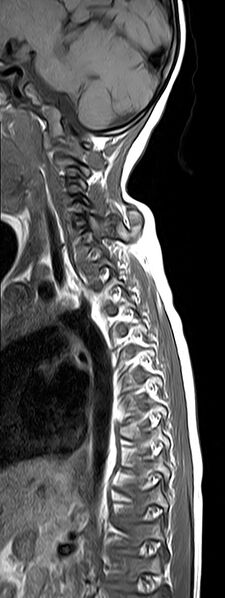
File:Bilateral Sprengel deformity with Klippel-Feil syndrome (Radiopaedia 66395-75650 Sagittal T1 9).jpg
Jump to navigation
Jump to search

Size of this preview: 225 × 598 pixels. Other resolutions: 90 × 240 pixels | 384 × 1,020 pixels.
Original file (384 × 1,020 pixels, file size: 175 KB, MIME type: image/jpeg)
Summary:
| Description |
|
| Date | Published: 19th Feb 2019 |
| Source | https://radiopaedia.org/cases/bilateral-sprengel-deformity-with-klippel-feil-syndrome |
| Author | Dr Ammar Haouimi |
| Permission (Permission-reusing-text) |
http://creativecommons.org/licenses/by-nc-sa/3.0/ |
Licensing:
Attribution-NonCommercial-ShareAlike 3.0 Unported (CC BY-NC-SA 3.0)
File history
Click on a date/time to view the file as it appeared at that time.
| Date/Time | Thumbnail | Dimensions | User | Comment | |
|---|---|---|---|---|---|
| current | 06:13, 14 June 2021 | 384 × 1,020 (175 KB) | Fæ (talk | contribs) | Radiopaedia project rID:66395 (batch #4331-9 A9) |
You cannot overwrite this file.
File usage
There are no pages that use this file.