File:Bilateral adrenal myelolipoma (Radiopaedia 63058-71537 Coronal T1 fat sat 1).jpg

From NC Commons
Jump to navigation Jump to search

Bilateral_adrenal_myelolipoma_(Radiopaedia_63058-71537_Coronal_T1_fat_sat_1).jpg(216 × 288 pixels, file size: 4 KB, MIME type: image/jpeg)

Summary:

Description
  • Radiopaedia case ID: 63058
  • Image ID: 42434032
  • Image stack position: 1/104
  • Plane projection: Coronal
  • Aux modality: T1 fat sat
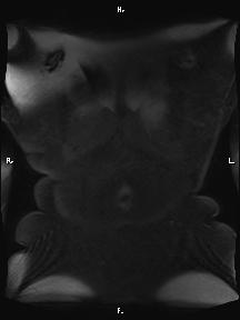
  • Study findings: A large well defined heterosignal mass is seen at right suprarenal region. The mass seems to be originating from right adrenal gland and causing inferior displacement of the right kidney. The mass contains areas with fat and soft tissue signal intensity. After contrast injection, nonfatty components of the mass show heterogeneous enhancement. The mass has pressure effect on the IVC but there is no sign of vascular invasion. Another small similar mass is seen in the left adrenal gland too.
  • Modality: MRI
  • System: Urogenital
  • Findings: A large well-defined fat-ontaining mass lesion was noted in the right adrenal gland. Soft tissue elements (myeloid components) were noted in the tumoral mass. After IV contrast media injection, mild enhancement is evident at these areas. The right kidney is displaced posteroinferiorly . Local invasion was not evident. Another smaller fat containing lesion was seen in the left adrenal gland.
Date Published: 12th Sep 2018
Source https://radiopaedia.org/cases/bilateral-adrenal-myelolipoma-1
Author Mohammad Taghi Niknejad
Permission
(Permission-reusing-text)
http://creativecommons.org/licenses/by-nc-sa/3.0/

Licensing:

Attribution-NonCommercial-ShareAlike 3.0 Unported (CC BY-NC-SA 3.0)

File history

Click on a date/time to view the file as it appeared at that time.

Date/TimeThumbnailDimensionsUserComment
current06:11, 11 June 2021Thumbnail for version as of 06:11, 11 June 2021216 × 288 (4 KB) (talk | contribs)Radiopaedia project rID:63058 (batch #4051-116 E1)