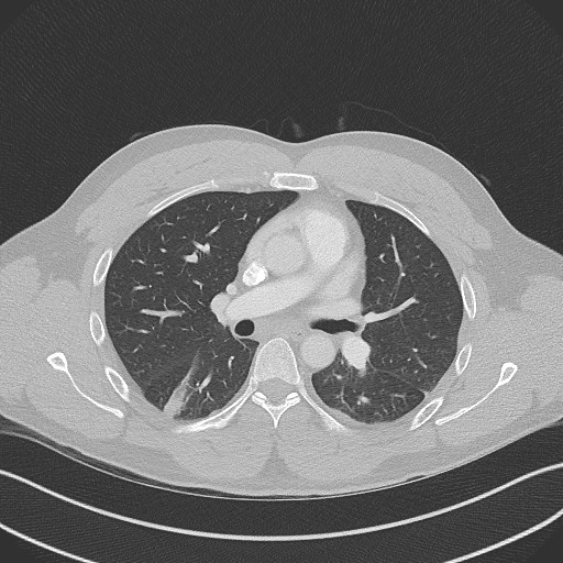
File:Bilateral atelectasis (Radiopaedia 48666-53675 Axial lung window 52).jpg
Jump to navigation
Jump to search
No higher resolution available.
Bilateral_atelectasis_(Radiopaedia_48666-53675_Axial_lung_window_52).jpg (512 × 512 pixels, file size: 78 KB, MIME type: image/jpeg)
Summary:
| Description |
|
| Date | Published: 20th Oct 2016 |
| Source | https://radiopaedia.org/cases/bilateral-atelectasis |
| Author | Andrew Murphy |
| Permission (Permission-reusing-text) |
http://creativecommons.org/licenses/by-nc-sa/3.0/ |
Licensing:
Attribution-NonCommercial-ShareAlike 3.0 Unported (CC BY-NC-SA 3.0)
File history
Click on a date/time to view the file as it appeared at that time.
| Date/Time | Thumbnail | Dimensions | User | Comment | |
|---|---|---|---|---|---|
| current | 09:44, 11 June 2021 | | 512 × 512 (78 KB) | Fæ (talk | contribs) | Radiopaedia project rID:48666 (batch #4064-52 A52) |
You cannot overwrite this file.
File usage
The following 2 pages use this file:
.jpg&oldid=516848)