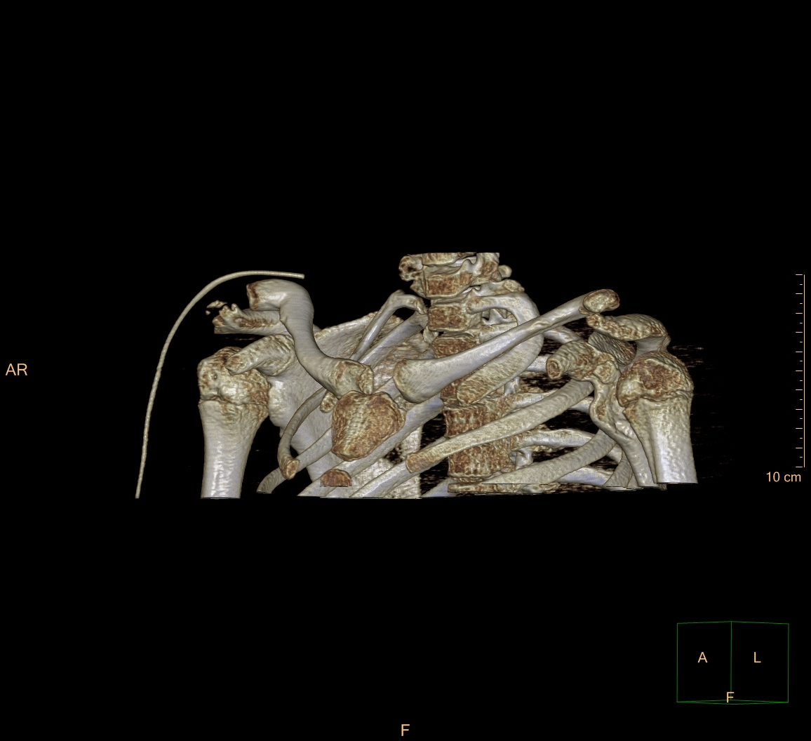
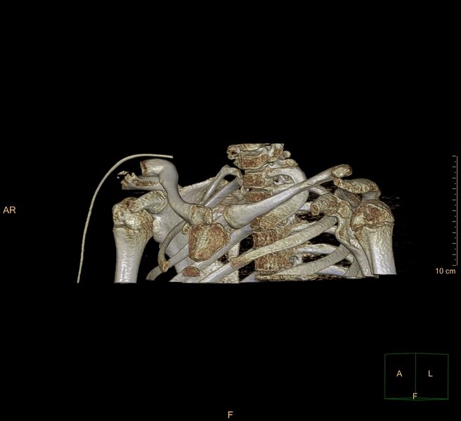
File:Bilateral chronic posterior shoulder dislocation (Radiopaedia 39213-41452 D 33).jpg
Jump to navigation
Jump to search
Size of this preview: 656 × 599 pixels. Other resolutions: 263 × 240 pixels | 526 × 480 pixels | 841 × 768 pixels | 1,156 × 1,056 pixels.
Original file (1,156 × 1,056 pixels, file size: 254 KB, MIME type: image/jpeg)
Summary:
| Description |
|
| Date | Published: 3rd Sep 2016 |
| Source | https://radiopaedia.org/cases/bilateral-chronic-posterior-shoulder-dislocation |
| Author | Stan Buckens |
| Permission (Permission-reusing-text) |
http://creativecommons.org/licenses/by-nc-sa/3.0/ |
Licensing:
Attribution-NonCommercial-ShareAlike 3.0 Unported (CC BY-NC-SA 3.0)
File history
Click on a date/time to view the file as it appeared at that time.
| Date/Time | Thumbnail | Dimensions | User | Comment | |
|---|---|---|---|---|---|
| current | 22:32, 11 June 2021 | | 1,156 × 1,056 (254 KB) | Fæ (talk | contribs) | Radiopaedia project rID:39213 (batch #4108-129 D33) |
You cannot overwrite this file.
File usage
There are no pages that use this file.
.jpg&oldid=521550)