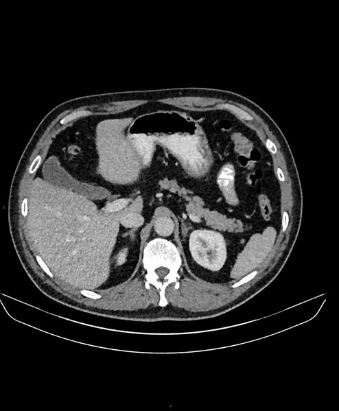
File:Bilateral duplex collecting systems (Radiopaedia 60831-68608 A 22).jpg
Jump to navigation
Jump to search

Size of this preview: 494 × 599 pixels. Other resolutions: 198 × 240 pixels | 396 × 480 pixels | 633 × 768 pixels | 844 × 1,024 pixels | 1,504 × 1,824 pixels.
Original file (1,504 × 1,824 pixels, file size: 246 KB, MIME type: image/jpeg)
Summary:
| Description |
|
| Date | Published: 4th Jun 2018 |
| Source | https://radiopaedia.org/cases/bilateral-duplex-collecting-systems-1 |
| Author | Derek Smith |
| Permission (Permission-reusing-text) |
http://creativecommons.org/licenses/by-nc-sa/3.0/ |
Licensing:
Attribution-NonCommercial-ShareAlike 3.0 Unported (CC BY-NC-SA 3.0)
File history
Click on a date/time to view the file as it appeared at that time.
| Date/Time | Thumbnail | Dimensions | User | Comment | |
|---|---|---|---|---|---|
| current | 02:27, 12 June 2021 |  | 1,504 × 1,824 (246 KB) | Fæ (talk | contribs) | Radiopaedia project rID:60831 (batch #4138-22 A22) |
You cannot overwrite this file.
File usage
The following page uses this file: