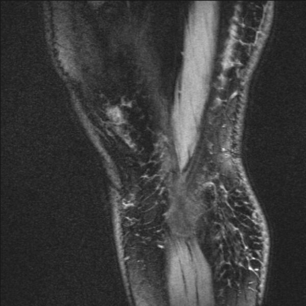
File:Bilateral focal pigmented villonodular synovitis (Radiopaedia 67643-77073 Sagittal T1 vibe 4).jpg
Jump to navigation
Jump to search

Size of this preview: 600 × 600 pixels. Other resolutions: 240 × 240 pixels | 618 × 618 pixels.
Original file (618 × 618 pixels, file size: 180 KB, MIME type: image/jpeg)
Summary:
| Description |
|
| Date | Published: 12th May 2019 |
| Source | https://radiopaedia.org/cases/bilateral-focal-pigmented-villonodular-synovitis |
| Author | Mostafa El-Feky |
| Permission (Permission-reusing-text) |
http://creativecommons.org/licenses/by-nc-sa/3.0/ |
Licensing:
Attribution-NonCommercial-ShareAlike 3.0 Unported (CC BY-NC-SA 3.0)
File history
Click on a date/time to view the file as it appeared at that time.
| Date/Time | Thumbnail | Dimensions | User | Comment | |
|---|---|---|---|---|---|
| current | 09:32, 12 June 2021 |  | 618 × 618 (180 KB) | Fæ (talk | contribs) | Radiopaedia project rID:67643 (batch #4164-148 G4) |
You cannot overwrite this file.
File usage
The following page uses this file: