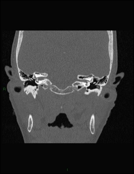
File:Bilateral frontal mucoceles (Radiopaedia 82352-96454 Coronal 301).jpg
Jump to navigation
Jump to search

Size of this preview: 462 × 599 pixels. Other resolutions: 185 × 240 pixels | 512 × 664 pixels.
Original file (512 × 664 pixels, file size: 45 KB, MIME type: image/jpeg)
Summary:
| Description |
|
| Date | Published: 23rd Sep 2020 |
| Source | https://radiopaedia.org/cases/bilateral-frontal-mucoceles |
| Author | Ian Bickle |
| Permission (Permission-reusing-text) |
http://creativecommons.org/licenses/by-nc-sa/3.0/ |
Licensing:
Attribution-NonCommercial-ShareAlike 3.0 Unported (CC BY-NC-SA 3.0)
File history
Click on a date/time to view the file as it appeared at that time.
| Date/Time | Thumbnail | Dimensions | User | Comment | |
|---|---|---|---|---|---|
| current | 12:46, 12 June 2021 |  | 512 × 664 (45 KB) | Fæ (talk | contribs) | Radiopaedia project rID:82352 (batch #4167-616 B301) |
You cannot overwrite this file.
File usage
The following page uses this file: