File:Bilateral locked facets (Radiopaedia 33850-35023 Sagittal bone window 53).png

From NC Commons
Jump to navigation Jump to search

Bilateral_locked_facets_(Radiopaedia_33850-35023_Sagittal_bone_window_53).png(512 × 512 pixels, file size: 117 KB, MIME type: image/png)

Summary:

Description
  • Radiopaedia case ID: 33850
  • Image ID: 10663606
  • Image stack position: 53/58
  • Plane projection: Sagittal
  • Aux modality: bone window
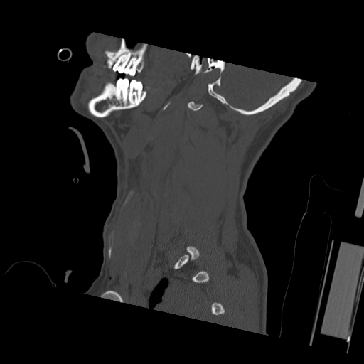
  • Study findings: Anterior discectomy and fusion is demonstrated at the level of C5/6. There is mild kyphosis at this level with mild retrolisthesis of C6 on C5. On the right, the facet joint is still dislocated and there is a 6 mm separation between the superior and inferior articular processes on the left.
  • Modality: CT
  • System: Musculoskeletal
  • Findings: There is approximately 50% subluxation of C5 on C6, with intervertebral disc space narrowing. This is associated with bilateral dislocated facets joints at this level, in association with abnormal widening of the interspinous space. No fracture is identified. There is severe central canal stenosis at this level, with significant impingement of the cervical cord. There is abnormal anterior vertebral soft tissue swelling centered at this level. Conclusion The findings are consistent with a severe hyperflexion injury, with bilateral locked facets at the C5/C6 level.
Date Published: 27th Jan 2015
Source https://radiopaedia.org/cases/bilateral-locked-facets-2
Author RMH Core Conditions
Permission
(Permission-reusing-text)
http://creativecommons.org/licenses/by-nc-sa/3.0/

Licensing:

Attribution-NonCommercial-ShareAlike 3.0 Unported (CC BY-NC-SA 3.0)

File history

Click on a date/time to view the file as it appeared at that time.

Date/TimeThumbnailDimensionsUserComment
current18:50, 12 June 2021Thumbnail for version as of 18:50, 12 June 2021512 × 512 (117 KB) (talk | contribs)Radiopaedia project rID:33850 (batch #4207-53 A53)

Metadata