File:Bilateral ossified cephalohematomas (Radiopaedia 35830).jpg
Jump to navigation
Jump to search
No higher resolution available.
Bilateral_ossified_cephalohematomas_(Radiopaedia_35830).jpg (513 × 513 pixels, file size: 45 KB, MIME type: image/jpeg)
Summary:
- Radiopaedia case ID: 35830
- Image ID: 193
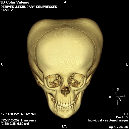
- Modality: CT
- System: Head & Neck
- Findings: There are bilateral bony protruberances from the parietal bones, which are seen to be due to thickened calvarium on the source images. This is the typical appearance of ossified cephalohematomas.
- Published: 1st May 2015
- Source: https://radiopaedia.org/cases/bilateral-ossified-cephalohaematomas
- Author: Laughlin Dawes
- Permission: http://creativecommons.org/licenses/by-nc-sa/3.0/
Licensing:
Attribution-NonCommercial-ShareAlike 3.0 Unported (CC BY-NC-SA 3.0)
File history
Click on a date/time to view the file as it appeared at that time.
| Date/Time | Thumbnail | Dimensions | User | Comment | |
|---|---|---|---|---|---|
| current | 18:28, 19 March 2021 | | 513 × 513 (45 KB) | Fæ (talk | contribs) | Radiopaedia project rID:35830 (batch #4117) |
You cannot overwrite this file.
File usage
The following page uses this file:
.jpg&oldid=8859611)