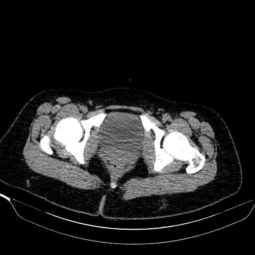
File:Bilateral ovarian dermoid (Radiopaedia 45714-49907 Axial non-contrast 43).JPEG
Jump to navigation
Jump to search
No higher resolution available.
Bilateral_ovarian_dermoid_(Radiopaedia_45714-49907_Axial_non-contrast_43).JPEG (512 × 512 pixels, file size: 35 KB, MIME type: image/jpeg)
Summary:
| Description |
|
| Date | Published: 5th Jun 2016 |
| Source | https://radiopaedia.org/cases/bilateral-ovarian-dermoid |
| Author | Aliakbar Sahraei |
| Permission (Permission-reusing-text) |
http://creativecommons.org/licenses/by-nc-sa/3.0/ |
Licensing:
Attribution-NonCommercial-ShareAlike 3.0 Unported (CC BY-NC-SA 3.0)
File history
Click on a date/time to view the file as it appeared at that time.
| Date/Time | Thumbnail | Dimensions | User | Comment | |
|---|---|---|---|---|---|
| current | 03:21, 13 June 2021 | | 512 × 512 (35 KB) | Fæ (talk | contribs) | Radiopaedia project rID:45714 (batch #4248-43 A43) |
You cannot overwrite this file.
File usage
There are no pages that use this file.
.JPEG&oldid=532568)