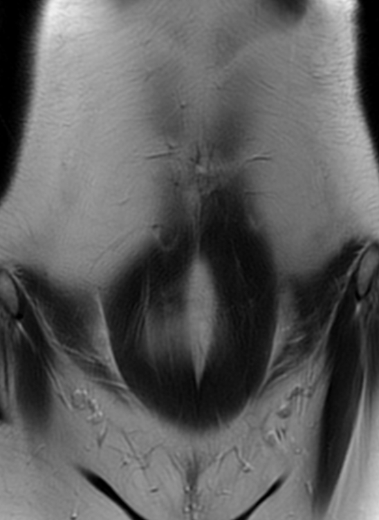
File:Bilateral ovarian fibrothecomas - adolescent (Radiopaedia 86604-102707 Coronal T2 6).jpg
Jump to navigation
Jump to search
Size of this preview: 436 × 599 pixels. Other resolutions: 175 × 240 pixels | 548 × 753 pixels.
Original file (548 × 753 pixels, file size: 129 KB, MIME type: image/jpeg)
Summary:
| Description |
|
| Date | Published: 8th Jun 2021 |
| Source | https://radiopaedia.org/cases/bilateral-ovarian-fibrothecomas-adolescent |
| Author | Dr Ammar Haouimi |
| Permission (Permission-reusing-text) |
http://creativecommons.org/licenses/by-nc-sa/3.0/ |
Licensing:
Attribution-NonCommercial-ShareAlike 3.0 Unported (CC BY-NC-SA 3.0)
File history
Click on a date/time to view the file as it appeared at that time.
| Date/Time | Thumbnail | Dimensions | User | Comment | |
|---|---|---|---|---|---|
| current | 06:15, 13 June 2021 | | 548 × 753 (129 KB) | Fæ (talk | contribs) | Radiopaedia project rID:86604 (batch #4255-108 D6) |
You cannot overwrite this file.
File usage
There are no pages that use this file.
.jpg&oldid=533724)