File:Bilateral perched facets with cord injury (Radiopaedia 45587-49714 Sagittal STIR 13).jpg

From NC Commons
Jump to navigation Jump to search

Bilateral_perched_facets_with_cord_injury_(Radiopaedia_45587-49714_Sagittal_STIR_13).jpg(512 × 512 pixels, file size: 117 KB, MIME type: image/jpeg)

Summary:

Description
  • Radiopaedia case ID: 45587
  • Image ID: 22906892
  • Image stack position: 13/16
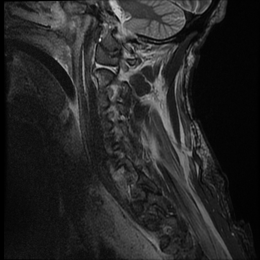
  • Plane projection: Sagittal
  • Aux modality: STIR
  • Study findings: Moderate anterior epidural hematoma extending from the clivus fracture to C7, with a maximal thickness of 6 mm, abutting the cord throughout its length. Small amount of posterior epidural hematoma at C6/7.  Incompletely imaged, likely small volume posterior epidural hematoma at T2-3 level. C6-7 discoligamentous rupture with bilateral perched facets and 8 mm anterior displacement of C6 on C7. Disruption of the ALL, PLL and ligamentum flavum, as well as the interspinous ligaments. Disc material/hemorrhage posterior to C6 abuts the cord. The cord is compressed at this level, with evidence of cord hemorrhage.  The abnormal cord signal extends to the C7-T1 level. The rest of the cord has normal signal. Right C2 lateral mass fracture extending into the odontoid process.  Fluid within the right C1-2 lateral articulation, with mild widening of the joint. Clivus and occipital condyle fractures, with tearing of the tectorial membrane at the clivus fracture site.  The apical ligament is torn.  The alar ligaments are intact, with sprain of the left alar ligament. C7 anterosuperior corner avulsion fracture fragment, with edema along the superior body. Left C1 lateral mass fracture extending into the foramen transversarium. T1 vertebral body rounded to T2 hyperintense lesion, with faint T1 hyperintensity in keeping with hemangioma. Vertebral artery flow voids are maintained throughout. Large amount of posterior interspinous edema.
  • Modality: MRI
  • System: Trauma
  • Findings: Fracture through the left lateral mass of C1 which involves the foramen transversarium. Fracture through the right lateral mass of C2 which involves the foramen transversarium. Fracture through the right C5 transverse process.  Fracture through the C5 spinous process. Fracture dislocation at C6-7. Fracture through the anteroinferior corner of the C6 vertebral body. Fracture through the right C6 pedicle extending to the foramen transversarium.  Fracture through the C6 laminae bilaterally and spinous process. Traumatic anterolisthesis of C6 on C7 by approximately 12 mm.  The left C6-7 facet joint is perched.  The right C6-7 facet joint is perched laterally but locked medially with small fracture fragments seen adjacent. Anterior epidural haematonma from C2 to C6-7, largest at C6-7.
Date Published: 31st May 2016
Source https://radiopaedia.org/cases/bilateral-perched-facets-with-cord-injury
Author Andrew Dixon
Permission
(Permission-reusing-text)
http://creativecommons.org/licenses/by-nc-sa/3.0/

Licensing:

Attribution-NonCommercial-ShareAlike 3.0 Unported (CC BY-NC-SA 3.0)

File history

Click on a date/time to view the file as it appeared at that time.

Date/TimeThumbnailDimensionsUserComment
current13:58, 13 June 2021Thumbnail for version as of 13:58, 13 June 2021512 × 512 (117 KB) (talk | contribs)Radiopaedia project rID:45587 (batch #4272-45 C13)

There are no pages that use this file.