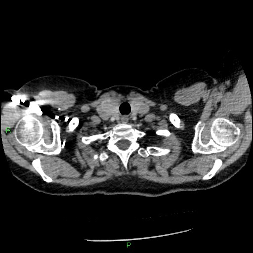
File:Bilateral pulmonary emboli (Radiopaedia 32700-33669 Axial C+ CTPA 5).jpg
Jump to navigation
Jump to search
Bilateral_pulmonary_emboli_(Radiopaedia_32700-33669_Axial_C+_CTPA_5).jpg (512 × 512 pixels, file size: 44 KB, MIME type: image/jpeg)
Summary:
| Description |
|
| Date | Published: 9th Dec 2014 |
| Source | https://radiopaedia.org/cases/bilateral-pulmonary-emboli |
| Author | Derek Smith |
| Permission (Permission-reusing-text) |
http://creativecommons.org/licenses/by-nc-sa/3.0/ |
Licensing:
Attribution-NonCommercial-ShareAlike 3.0 Unported (CC BY-NC-SA 3.0)
File history
Click on a date/time to view the file as it appeared at that time.
| Date/Time | Thumbnail | Dimensions | User | Comment | |
|---|---|---|---|---|---|
| current | 20:00, 13 June 2021 |  | 512 × 512 (44 KB) | Fæ (talk | contribs) | Radiopaedia project rID:32700 (batch #4302-5 A5) |
You cannot overwrite this file.
File usage
The following page uses this file:
