File:Bilateral skin folds mimicking pneumothoraces (Radiopaedia 59736).png
Jump to navigation
Jump to search

Size of this preview: 463 × 599 pixels. Other resolutions: 185 × 240 pixels | 371 × 480 pixels | 594 × 768 pixels | 792 × 1,024 pixels | 1,534 × 1,984 pixels.
Original file (1,534 × 1,984 pixels, file size: 1.88 MB, MIME type: image/png)
Summary:
- Radiopaedia case ID: 59736
- Image ID: 37942554
- Modality: X-ray
- System: Chest
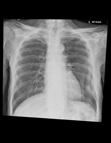
- Findings: AP erect film. Heart is not enlarged. Lungs and pleural spaces clear. On the left are two vertical lines paralleling the lateral chest wall and on the right a single line. These appearances are consistent with prominent bilateral skin folds which may mimic lung edges as seen with pneumothoraces. Lung markings are clearly visible peripheral to the skin folds. No subcutaneous emphysema. No rib fractures. No evidence of hemothorax. Bilateral degenerative disease of the ACJs and glenohumeral joints.
- Published: 22nd Apr 2018
- Source: https://radiopaedia.org/cases/bilateral-skin-folds-mimicking-pneumothoraces
- Author: Daniel J Bell
- Permission: http://creativecommons.org/licenses/by-nc-sa/3.0/
Licensing:
Attribution-NonCommercial-ShareAlike 3.0 Unported (CC BY-NC-SA 3.0)
File history
Click on a date/time to view the file as it appeared at that time.
| Date/Time | Thumbnail | Dimensions | User | Comment | |
|---|---|---|---|---|---|
| current | 18:44, 19 March 2021 | | 1,534 × 1,984 (1.88 MB) | Fæ (talk | contribs) | Radiopaedia project rID:59736 (batch #4196) |
You cannot overwrite this file.
File usage
There are no pages that use this file.
.png&oldid=8859600)