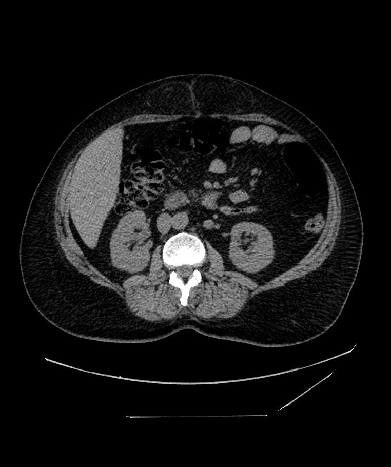
File:Bilateral sporadic synchronous clear cell renal cell carcinoma (Radiopaedia 85035-100573 Axial non-contrast 64).jpg
Jump to navigation
Jump to search
Size of this preview: 502 × 600 pixels. Other resolutions: 201 × 240 pixels | 402 × 480 pixels | 762 × 910 pixels.
Original file (762 × 910 pixels, file size: 99 KB, MIME type: image/jpeg)
Summary:
| Description |
|
| Date | Published: 22nd Dec 2020 |
| Source | https://radiopaedia.org/cases/bilateral-sporadic-synchronous-clear-cell-renal-cell-carcinoma |
| Author | Ammar Ashraf |
| Permission (Permission-reusing-text) |
http://creativecommons.org/licenses/by-nc-sa/3.0/ |
Licensing:
Attribution-NonCommercial-ShareAlike 3.0 Unported (CC BY-NC-SA 3.0)
File history
Click on a date/time to view the file as it appeared at that time.
| Date/Time | Thumbnail | Dimensions | User | Comment | |
|---|---|---|---|---|---|
| current | 02:03, 14 June 2021 | | 762 × 910 (99 KB) | Fæ (talk | contribs) | Radiopaedia project rID:85035 (batch #4329-64 A64) |
You cannot overwrite this file.
File usage
The following page uses this file:
.jpg&oldid=540956)